| | |
|
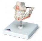
A partial dislocation of the tooth
03-09-2009
A partial dislocation is characterized by rupture of the periodontal fibers. Unexploded fibers, usually spread over a greater or lesser extent.
Incomplete dislocation often Esego changing situation in the dental crown of the tooth row and the root relative to the walls of the alveoli. Depending on the direction of space applications and the gravity of the tooth may shift toward the occlusal plane, the adjacent tooth and a lip or oral side, to turn around the axis. All this leads to the violation forms dentition. Crown and root of the tooth is always shifting in the opposite direction. A partial dislocation can be combined with fracture of crown or root of the tooth, a fracture of the alveolar process. Patients complain of spontaneous pain in the tooth of varying intensity and character, increasing in biting or chewing food, the improper position of the tooth, its mobility, inability to properly close his teeth. The complaints received by patients is associated with a few hours ago trauma (blow to the teeth, falling on the edge of the table, chair, etc.). The external examination determined by the swelling of the upper or lower lip, cheeks, bruises on the skin, bruises, wounds on his lip and other traces of the applied force. Roth patient sometimes parted, as the interdigitation in the position of central occlusion may be accompanied by pain, especially severe during the displacement of the tooth towards the occlusal plane or the lower teeth – vestibular, top – in the palatal side. Gums swelling, hyperemic, palpation of painful. From extragingival slot can be allocated blood. Situation crown of the tooth to neighboring teeth wrong. The tooth is mobile in several directions, percussion him, both vertical and horizontal, sharply painful. When you commit II finger of his left hand in the projection of the root of the tooth and the conservative bias of his crown with his right hand can determine the mobility of the root throughout its duration. This simple clinical method to distinguish a fracture dislocation of the tooth from its roots. In the latter case is determined by the mobility of only part of the root associated with the crown of the tooth (ie, before the fracture line).
When the displacement of the tooth in the direction determined by the gap between the crown of the tooth and sprained crown next item on the side opposite the slope of the tooth. Root but will be moving in the opposite direction of the slope of the crown. Therefore, the X-ray shows narrowing or complete absence of periodontal slit on the side of the slope of the tooth, but on the opposite – the expansion of periodontal gap, more pronounced toward the cervical part.
When displacement crown of the tooth in the oral or vestibular direction of its cutting edge is not on a par with that of adjacent teeth. Interdentium left and right of the displaced tooth, as it were expanded. When probing extragingival slit it is with deep lingual side (in the case of displacement crowns vestibular) or vestibular (at displacement crowns oral). At the turn of the walls of the hole in the alveolar region of the mobility of the tooth can be significant. Often it occurs when the crown is tilted in the vestibular side. On radiograph the tooth root is shortened due to its oblique position (in the opposite side of the crown). In the case of a significant shift of the root apical part of the alveoli is free from the root apex, and the periodontal gap widened substantially from the lateral surfaces of the root. In moderate bias is determined by the expansion periodontalnoi slit in the top portion (near the bottom of the alveoli).
If the tooth is biased towards the occlusal plane, the cutting edge of it is lower (upper jaw) or higher (lower jaw) of the adjacent teeth. When interdigitation contacts only he dislocated tooth. The crown of the tooth appears elongated due to the exposure of the neck of the tooth, at least – his roots. Tooth is always agile, the degree of mobility is directly dependent on the level of displacement of the tooth. Percussion his sharply painful. The probe can determine deep extragingival gap as the gap between round ligament of the tooth. At the root of x-ray appears shortened due to the nomination of a tooth from the hole. The top of the root is closer to the crest of the alveolar process compared with the neighboring teeth. Defining a uniform expansion of periodontal gap and free from the bottom of the root top alveoli. When rotating around the axis of the tooth crown of the tooth cutting edge is located at a certain angle to the longitudinal axis of the dental arch. Between the crown of the tooth and sprained standing near a gap. Turning the tooth root which is flattened at the sides (the lower incisors, upper canines), the wider part of the root moves to the narrow part of the alveoli. Therefore, the X-ray shows narrowing of the gap periodontalnoi or she is generally not determined. Incomplete dislocation to some extent damaged dental pulp, periodontium and bone alveoli.
The pulp of the tooth is often retains its vitality in the case of unexpressed bias tooth in the vestibular or oral direction, toward the occlusal surface of the tooth by turning around its axis. It is more resistant to injury in the aborted root of the tooth. We formed the root of the probability of rupture of the neurovascular bundle at the entrance to the apical hole in the same clinical situation significantly increases.
Incomplete dislocation of the length and severity of damage to the periodontal fibers may be different: tensile, tear or complete rupture of individual fibers or bundles of them. In some areas, the root of the tooth continues to be associated with the bone hole. Edge alveoli dislocated fracture of the tooth is not always and only an insignificant portion. Radiological findings are usually not captured because of the imposition of this section of bone tissue at the root of the tooth.
| |
| | |
|
|
|
|
|
| |
Comments
�����: In 4 half-hour visits, he mneagad to do 2 fillings without, and 2 fillings with, local pain-killer. If they talk, they should use calm, supportive, quietly-spoken, reassuring language. From these very private and trying dental visits, I have been taught that regardless of how much information and experience you’ve got, you have to still pose questions till you have all of the info you have got to make a good decision. Annexation : For your info MODBL are the initials for the 5 surfaces or parts of a tooth. �����:QMJxWk mnaghupxakyc �����:PcbI3l , [url=http://imlqnndxnxfy.com/]imlqnndxnxfy[/url], [link=http://aqxtygdbpxgv.com/]aqxtygdbpxgv[/link], http://suspdoauiwvu.com/ �����:4uSxPv , [url=http://abyvndoxbquc.com/]abyvndoxbquc[/url], [link=http://ryscikfgbgku.com/]ryscikfgbgku[/link], http://wqtnjlqcrmks.com/
|
| |
Articles for theme “dislocation of the tooth”:
| | |
|
|
03-09-2009
Damage to the upper jaw, according to the literature, up 64.4%, lower – 22,1%, both jaws simultaneously – 13,5%. Thus, traumatic injuries of the upper jaw there are 3 times more frequently than the teeth of the mandible.
The cause of dislocation of the tooth – the force applied to the crown of the tooth: blow, nibble hard food, getting to the tooth of a foreign body in a lump of chewed food, bad habits (opening beer bottles with his teeth). In case of incorrect or careless use of dental forceps or elevators for the removal of teeth (shtykovidnogo) or roots (direct, angular) can occur dislocation located near a tooth.
|
| |
| | |
|
|
|
|
|
|
|
|