|
|
|
|
|
03-09-2009
For convenience of presentation, suppose that the left fissure fracture runs along the upper type, and the right – on average. The line is a fracture in the frontal connections sprouts upper jaw with the nasal part of frontal bone. Left it extends characteristic of Le Fort I (ris.3.12), and the right – for the Le Fort II (see the relevant section, as outlined above). Patients may complain of pain in the upper jaw, with the increasing interdigitation or attempted biting or chewing food, improper closing of the teeth, foreign body sensation in the throat, choke and retch, insufficient opening of the mouth, double vision.
|
|
|
|
|
|
|
|
|
|
|
|
|
|
|
03-09-2009
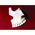
Fracture of the upper jaw of Le Fort III (bottom type) Fracture line passes through the edge of pear-shaped holes, backwards and slightly above the bottom of the maxillary sinus. It crosses skuloalveolyarny ridge, goes through the hill of the upper jaw and extends to the lower third of the pterygium sprouts sphenoid bone (see 3.10). Sometimes pterygoid bone is not broken off with the upper jaw, and is separated from her mound at their place of seam. In these cases, pressure on the hook pterygium the process, as described above, is not accompanied by pain and may complicate diagnosis.
|
|
|
|
|
|
|
|
|
|
|
|
|
|
|
03-09-2009
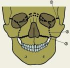
Fracture line passes through the place of the frontal sprouts upper jaw with the nasal part of frontal bone on the inner wall of the orbit to the infraorbital fissure. Further extends anteriorly on the lower wall of the orbit to the infraorbital region, crossing his or skuloverhnechelyust Board the seam, or close to it. Then comes down and backwards on the front surface of the upper jaw, extending to the pterygoid bone sphenoid bone (sometimes on the border of the upper and middle third) (ris.3.7).
|
|
|
|
|
|
|
|
|
|
|
|
|
|
|
03-09-2009
The line is a fracture at the junction of the frontal sprouts upper jaw with the nasal part of frontal bone in her lattice notch. The front edge of the latter is connected to the nasal bones, and the rear – with the front edge of the plate perforated ethmoid bone, which is involved in the formation of the skull base in front of his pit. Rear bow sections of the frontal bone contains cells in contact with the ethmoid bone and forming the roof of its cells. Then the fracture line passes through the inner wall of the eye socket to the junction of the upper-and infraorbital slits, passes to the outer wall of the orbit, extending to her up and anterior to verhnenaruzhnogo its corner.
|
|
|
|
|
|
|
|
|
|
|
|
|
|
|
03-09-2009
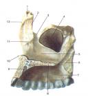
Land increased strength of the upper jaw depending on the structure of bone tissue related to its formation in phylogenesis. Strong space correspond to points of ossification, the weak – the intermediate lines. Plots of reduced strength are placed along the joints connecting it with the other bones of the facial skeleton and the bones forming the base of the skull. These sites often pass through neognestrelnogo fracture of the upper jaw. This may explain the fact (along with other factors – venue traumatic force, its direction relative to the buttresses), which is most often a fracture line is not strictly within the anatomical boundaries of the upper jaw, but extends to the neighbor, the associated bone.
|
|
|
|
|
|
|
|
|
|
|
|
|
|
|
03-09-2009
Sphenoid bone (os sphenoidale) is located in the middle of the skull base. It includes body and three pairs of appendages: small and large wings, and pterygoid processes. The body is the middle part of the bone. It is wedge-shaped atrium, covered with mucous membrane and filled with air. The upper surface of the body, facing into the cavity of the skull, is in the middle of deepening – Turkish saddle. Small wings of sphenoid bone away from the body on each side of the two roots. Between them is the visual channel, through which pass the optic nerve and orbital artery.
|
|
|
|
|
|
|
|
|
|
|
|
|
|
|
03-09-2009
Ethmoid bone (os etmoidale) consists of the middle part and two lateral. The middle part is a small horizontal lattice plate (lamina cribrosa) and a large perpendicular (lamina perpendicularis). Lateral parts – a large number of pneumatic cells bounded by thin lamellae and forming a latticed labyrinth. The cells are covered with mucous membrane. Ethmoid bone is located in the lattice cutting out the frontal bone. Lattice plate it is part of the cerebral skull. The remaining parts are involved in the formation of the skeleton of the nasal cavity and the inner wall of the orbit.
|
|
|
|
|
|
|
|
|
|
|
|
|
|
|
03-09-2009
Frontal bone (os frontale) is involved in the formation of the roof of the skull base, the walls of the orbit, the nasal cavity. In the frontal bone distinguish frontal scales, bow, and The orbital part. In scales distinguish the two surfaces: the external and internal. The outer surface is convex, has 2 frontal tuber and beneath crescent-shaped rolls – eyebrows. Between the frontal tubercles and eyebrows formed playground – intercilium (glabella). Lateral located zygomatic process, which are connected with the zygomatic bone.
|
|
|
|
|
|
|
|
|
|
|
|
|
|
|
03-09-2009
Nasal bone (os nasale) – doubles the bone, forming the root and part of the back of the nose. Medial margin of nasal bone is connected to the opposite bone, lateral – with frontal offshoot of the upper jaw, upper – with a nasal edge of the frontal bone. The lower edge of the upper limit of the pear-shaped openings. The inner surface has a lattice furrow, which is located anterior ethmoid nerve and 1-2 small nasal openings, which open on the outer surface.
|
|
|
|
|
|
|
|
|
|
|
|
|
|
|
03-09-2009
Lacrimal (os lacrimale) – pair, the smallest and thinnest facial bones. Located in the anterior medial wall of the orbit between the frontal offshoot of the upper jaw and the orbital plate of ethmoid bone. The upper edge is connected to the nasal part of frontal bone, lower – the upper jaw; medial surface it adjoins to the ethmoid bone, covering the anterior end of the maze, and lateral – turned into the cavity of the eye socket. On the lateral surface of the vertically located posterior lacrimal crest, and anterior to it – lacrimal groove.
|
|
|
|
|
|
|
|
|
|
|
|
|
|
|
03-09-2009
Palatine bone (os palatinum) – doubles the bone, located between the upper jaw (front) and wing-spikes sphenoid (behind). Participates in the formation of the walls of the orbit, nose, mouth. It consists of two plates: horizontal (lamina horisontalis) and perpendicular (lamina peipendicularis). Horizontal plate is connected to the front edge of palatine offshoot of the upper jaw, forming the back of the hard palate. The rear edge of its limits hoany. Along the medial margin of the nasal surface is the nasal crest (crista nasalis), which joins shovel.
|
|
|
|
|
|
|
|
|
|
|
|
|
|
|
03-09-2009
Zygomatic bone (os zygomaticum) – doubles the bone, consisting of thick plates of a compact and a small layer of spongy substance. It distinguished cheek, orbital, temporal surface. Cheek surface (lateral) – convex, square shape, its anteroinferior edge in contact with the zygomatic process of maxilla. The upper part is a frontal processes, which connects with the zygomatic process of frontal bone, and its posterior part – with a large wing of the sphenoid bone. Inferolateral angle formed temporal appendage, which adjoins to the zygomatic process of temporal bone and forms together with the zygomatic arch.
|
|
|
|
|
|
|
|
|
|
|
|
|
|
|
03-09-2009
The upper jaw bone of steam, is connected with the zygomatic, frontal, nasal, Lattice, sphenoid, lacrimal bone. In her distinguished body and four sprouts: frontal, alveolar, palatal and zygomatic. In the body of the upper jaw is aeriferous maxillary sinus, the walls of which are represented by thin lamellae of the compact substance. There are four body surface of the upper jaw: the front, infratemporal, orbital, nasal. The front surface, fades anterior, limited infraorbital edge (top), skuloalveolyarnym crest and the zygomatic process (laterally), alveolar bone (below), the nasal notch (medial).
|
|
|
|
|
|
|
|
|
|
|
|
|
|
|
03-09-2009
Under the conduction anesthesia is necessary reponirovat otlomok and install it in the correct position. In this case the fingers is fixed and the health section of the alveolar process in order to make sure we do not further break the mucous membrane and periosteum, which is essential for the outcome of treatment. Immobilization of fragments can be accomplished through a smooth tire-bracket, if the undamaged portion of the alveolar process has a sufficient number of stable teeth (at least 2-3 on each side of the line of fracture).
|
|
|
|
|
|
|
|
|
|
|
|
|
|
|
03-09-2009
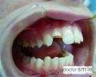
Isolated fracture of the alveolar process is the result of an inflection or shift in the place of application of force. Most widespread classification of fractures of the alveolar process, developed KS Sound (1968). According to this classification are the following types of fractures: ▲ partial – the line of fracture passes through the outer part of the alveolar process; fracture occurs outside of a compact disc within the wells of several teeth and part of the interdental partitions; ▲ part – the line of fracture in the form of cracks through the entire thickness of the alveolar process, capturing the outer and inner compact discs, sponge, displacement of fragments does not occur; ▲ complete – two vertical lines of fracture united by one horizontal and pass through the entire thickness of the alveolar process; ▲ comminuted – the line of fractures intersect in several directions; ▲ to the bone defect – separation of broken alveolar bone.
|
|
|
|
|
|
|
|
|
|
|
|
|
|
|
03-09-2009
Treatment of patients with fracture of the crown of the tooth is aimed at restoring the lost crown of the tooth tissue. In the case of fracture of the tooth at the cervical orthopedic methods of reconstructing the lost crown a more promising, if the fracture line passes over extragingival attachment (making pins tooth crowns Stump). Sometimes to implement the plan of orthopedic treatment appropriate excision of the edges of the mucous membrane. In the literature there are indications of the effectiveness of orthodontic preparation for orthopedic treatment.
|
|
|
|
|
|
|
|
|
|
|
|
|
|
|
03-09-2009
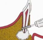
When incomplete dislocation of the tooth under conduction anesthesia should be carefully reponirovat it. At the same time his fingers have not only dislocated the tooth, but necessarily on neighboring and alveolar bone. It is necessary to prevent accidental breakdowns it in the opposite direction. The pressure on the tooth should be developed gradually and moderately, to not further damage the neurovascular bundle at the site of its entry in the apical hole. Proper conduct of repositioning can be defined by the absence of contact with the antagonist only reponiruemogo of the tooth.
|
|
|
|
|
|
|
|
|
|
|
|
|
|
|
03-09-2009
Fracture of the root of the tooth may occur near the neck of the tooth, in its central part, on the border of the middle and apical thirds, near the top. The direction of fracture lines often cross, rarely slightly oblique. It passed through the cement, the dentin, the tooth pulp. If the fracture line of two or more, to speak of comminuted fractures of the root. According NM Chupryninoy et al. (1993), the roots of incisors often break between the middle and apical third (69.0%), equally often – in the neck and middle (14.
|
|
|
|
|
|
|
|
|
|
|
|
|
|
|
03-09-2009
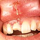
Fracture of crown of the tooth may occur within the enamel, which often breaks off crown angle; enamel and dentin with or without exposure of the outcrop koronkovoi pulp, enamel, dentine and cement – broke off all the bits in the cervical region along the enamel-dentinovoy border. With palatal side fabric cleave at an acute angle, with the vestibular – at right. The level of passing through the fracture determines the value of the Snap-Off crown and the ratio of the wound surface to pulpovoy chamber.
|
|
|
|
|
|
|
|
|
|
|
|
|
|
|
03-09-2009
Causes of tooth fracture are the same and cause dislocation. In addition, the crown of the upper teeth can be broken when removing the lower teeth with pliers, when conducting traction of the tooth, the doctor accidentally strikes a blow with the tongs in the teeth of the upper jaw. Fracture can occur in any part of the tooth. Chance broke off part of the crown without opening the cavity of the tooth and with the discovery of her, broke off all the crowns, the fracture of the root at a different level, change crowns and roots simultaneously.
|
|
|
|
|
|
|
|
|
|
|