Dental Pulp

06-11-2008
Dental Pulp
The dental pulp is the soft connective tissue that forms the inner core of each tooth. It is divided in the coronal part and the radicular part depending on its position. At the root tip, the radicular part of the pulp merges with the periodontal ligament. At this site, blood vessels and nerves enter the tooth. At the interface of pulp and dentin lies a continuous row of columnar odontoblasts. They are responsible for the deposition of dentinal matrix that may be either secondary as a physiologic process continuing as long as the tooth lives, or tertiary as a reply to some insult. In the latter situation, they lose their regular palissading architecture and may lie embedded in the matrix they have deposited. In the pulp, calcified particles may form, the so-called denticles. They will be discussed more extensively elsewhere During and after eruption, the tooth pierces through the oral mucosa while reduced enamel epithelium and oral epithelium coalesce. To maintain an adequate barrier between the inner and the outer environment, there has to be a farm connection between the epithelial lining of the oral cavity and the surface of the tooth. This requirement is fulfilled by the so-called junctional epithelium, a rim of specialised multilayered nonkeratinising squamous epithelium that forms a cuf attached to and surrounding the necks of the teeth. In a healthy situation, this epithelium is attached to the enamel surface, forming a tapering end at the border between root cementum and enamel. At its opposite end, the junctional epithelium ends in a free margin where it merges with the epithelial surface of the free gingiva, the part of the gingiva that is connected to the tooth surface. This latter epithelium is parakeratinised and shows extremely long and thin rete ridges that may split into several branches. Both the epithelial covering of the free gingiva as well as the junctional epithelium are supported by collagenous fbres fanning out from the root surface and connected to the root through embedding in the root-covering cementum.

Te junction between epithelium and tooth is very vulnerable as it depends only on adhesion between both surfaces. Slight infammatory alterations in the gingiva may already cause disruption of this attachment. Te consequences of this disruption will be discussed under the heading gingival and periodontal disease. Briefy, if there is loss of attachment of the junctional epithelium to the tooth surface, it may move downwards to lie adjacent to the root cementum and may establish a new attachment at that site.

Views: 2987 | Comments: 9 Send reply
 
Comments
Гость:
Barter Trade Swap ywood, fenceing pfreer four ft or higher wood or wire not picky, cheap mini van or smaller truck,electric gate, servalance cameras,black side by side frig, leather recliners, blond wood baby crib with matching dresser, king adjustable. Duel control bed, flat t.v., dental services teeth whiteing and implants, breast reconstruction , computer Or note pad, camper or RV, wii games, slot machine, weekend get away to??? Willing to barter carpet and installation. Or Vinyl. And installation send me an email of what you have to barter and what flooring. Needs you have we also have a thrift. Store so if you news something else and don’t need flooring let me know what you need and maybe we. Can barter Location: Colorado it’s NOT ok to contact this poster with services or other commercial interests

Гость:
Thanks for the info.Very interesting ineded. I learned about max protein intake in my fitness class. But i didn’t know it was as low as 7 grams/ hour. There is obviously variables included in this number, but the point is : 30 g protein bars = fat storage.What would you recommend for an athlete looking to build strength and conditioning? I am of fairly thin stature, 5 10 : 140 pounds : BMI = 21i dont put on weight easily, and i am not looking to. Rather, I want to gain strength and tone through exercises that use my own body weight as resistance. Given my financial position as a student, combined with my active lifestyle, I don’t receive enough protein daily.

Гость:
Hello Gary, After reading your post on your webtise about the heavy metals in protein shakes, I have a question. I have recently been in much want to gain more weight/muscle mass. I am 18 and have to high of a metabolism and I play highschool basketball and soon college. I have been taking precisionengineered muscle and weight gainer and had seen definite difference on the weight scale. I dont believe in the creatines. But it does say it has alot of amino acids and the calories,which I need more of, is 580. I am 124lbs. at the moment. Is this a harmful substance for my body?

Гость:
I started dinkring protein drinks at every meal with some oatmeal and flagseed for about a month and started having joint and Tendon problems (severe problems ) I was told by the doctor that it was because i was getting older and that i should not work out as much as i do ? I did not agree with that so i started to do some research on my diet since i figured that had to be it ? And found you post . thanks http://bwtvlyd.com [url=http://liiprb.com]liiprb[/url] [link=http://xlwgvv.com]xlwgvv[/link]

Гость:
Oh, that has been some bad luck for you, but I know, visiting detsnit is not my thing as well! 🙁 I hope it’ll go smoother than you expect! I’m already up this morning, since I have 2 tests + an exam at my university at the same time, you’ll be at the detsnit and these are on the most boring and hard to read subject ever. Quite unpleasant, too. I already keep fingers crossed for you!Hugs,Marina
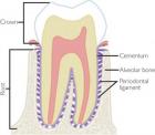
Гость:
Oh no! 🙁 is it tooth in the front?I have the exact same thing and I know how annoying it is to go and fix it when it berkas again!And of course i’ll be with you at 9! I never been afraid of the dentist cause since my childhood I have a fantastic dentist, she never hurt me and does an very good job! I go to her as I go to the hairdresser actually :)Love and Hugs

Гость:
I know that feel….That happened to me too. I neeedd to go to the dentist but he is too occupied so now I have a half tooth….Guess I’ll hve to wait to fix that for a loooooong time -.-I wish you a good luck tomorrow, even though the sound is annoying, think about the ones you love and all the positive stuff in life. It will be over soon for sure :)HugsEna :* http://rcoyebv.com [url=http://vvwyeh.com]vvwyeh[/url] [link=http://tvfqodfquik.com]tvfqodfquik[/link]

Гость:
Hej Anette!Samma sak he4nde min mor ff6r ne5gra me5ader sedan. Men hon gick inte till tandle4karen direkt, utan hon tog lite srpiulem och klistrade fast tanden igen 😀 Jaja, har man inte tid att ge5 till tandle4karen se5… Men hon har fixat tanden nu… tror jag…Men i alla fall, jag ska skicka lite positiv energi se5 att du klarar av den hemska upplevelsen :)Ha det bra!//Ellinor

Гость:
Hej Anette!Ne4r du le4ser det he4r se5 har du nog redan fixat tanden, se5 jag hopaps att allt gick bra :)Karin: Haha ke4nns bra att jag kunde fe5 ne5gon att skratta 😀 Superlim e4r ve4ldigt vanligt he4r hemma, och min mor anve4nder det inom sitt jobb ockse5. Vi ve4ntade nog bara pe5 att hon skulle gf6ra ne5got galet med det 😀 Ha det bra!//Ellinor http://ofrwylgt.com [url=http://cpujqh.com]cpujqh[/url] [link=http://xjibaz.com]xjibaz[/link]

Send reply
Name:

Text:

Pleas enter the code on picture:
Enter this code 

 
Articles for theme “Histology”:
Cementum
06-11-2008
Root cementum is visible as a tiny, slightly basophilic layer at the outer surface of the root part of the tooth and can be distinguished from dentin by the absence of tubuli. It obliterates the peripheral ends of the dentinal tubuli and lies adjacent to the mantle dentin. It is the product of fibroblasts from the dental follicle migrating to the root surface and differentiating into cementoblasts. Whether epithelial cells from Hertwig’s sheath contribute to initial cementum formation is controversial as has already been discussed.
Enamel
06-11-2008
Enamel content is almost 100% mineral, so it evades histologic evaluation in common paraffin sections from decalcified material. Only in immature tooth tissues, may it be seen in routinely prepared histologic slides due to a still higher proportion of organic components, where it is visible as a slightly basophilic the ready material. In this form, it may also form part of the histologic appearance of odontogenic tumours. Examination of mature enamel requires the employment of ground sections.
Histology
11-10-2008
Teeth consist for the major part of dentin. This material houses the dental pulp, the soft tissue core of the tooth consisting of myxoid connective tissue with blood vessels and nerves, and supports the enamel cap that covers the part of the tooth that is exposed to the oral cavity. In the root area, dentin is covered by cementum that fixes the collagenous fibres of the periodontal ligament onto the root surface. At the other side, these collagenus fibres are attached to the bone of the tooth socket and in this way, the tooth is fixed in  the jaw.
Dentin Histology of the Teeth
07-06-2008
Dentin is a specialized kind of bone formed by the odontoblasts but different in the sense that it does not contain complete cells but only cellular extensions, i.e., cytoplasmic extensions from the odontoblasts. These cross the full thickness of the dentin from the odontoblastic cell body that lies at the border between dentin and dental pulp to the junction between dentin and enamel. The tiny canals that house the odontoblastic extensions are recognizable as evenly spaced tubuli. This tubular nature is the histologic hallmark for dentin, not only in teeth but also in odontogenic lesions in which the nature of each mineralized material may not be recognizable at first sight.