1.1 Introduction
There are few scientific approaches to human identification that are more effective than a well-trained forensic dentist armed with a set of high-quality dental records and radiographs. Fingerprinting is probably the only other technique used with greater frequency, but as we know, the soft tissue of the extremities does not resist the ravages of time and environment like the enamel and dentin of human teeth. So, in terms of rapidity, degree of certainty, cost-fectiveness, and applicability to a wide range of intact, decomposing, or skeletonized remains, forensic odontology has been the identification method of choice.For the last two decades, however, a new science has appeared on the forensic stage and, as is the lot for many newcomers, forensic DNA analysis has been both celebrated for its extraordinary achievements and criticized for its complexity and disappointments. Miniscule amounts of biological evidence can be individualized and the results quantified using statistics so staggering that the courts and the public have come to expect the same sort of return on all types of forensic analyses. However, the process of DNA analysis is very slow in comparison to other forensic examinations, extremely expensive, and with few exceptions, must be conducted in highly specialized and fixed facilities. The very power of DNA and the ease with which large population databases can be developed have generated their own set of social problems and ethical concerns. With this new technology comes an increased risk to personal privacy that actually crosses generations, as well as the fear of genetic discrimination in employment and insurance sectors.
The traditional odontology community acknowledges the capabilities of DNA science as applied to human identification, bitemarks, and mass fatality management, and the dividing lines of the relationship are just beginning to become defined. Like the pathologist and the anthropologist, the odontologist’s area of expertise includes some of the DNA scientist’s favorite targets for analysis. The forensic odontologist will find himself called upon to select and prepare samples for submission to the laboratory and plan for disaster responses that will inevitably include forensic DNA support.
1.2 Molecular Biology and Inheritance
To fully appreciate the power of discrimination offered by the DNA molecule, one must first take a brief moment to grasp its simple elegance. Although earlier investigators initially suspected that proteins were actually the carriers of hereditary information, William Thomas Astbury demonstrated in 1944 that DNA is the sole genetic material of life.
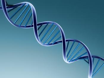
Although he received only minor recognition for his discovery, his successors became much more famous. James Watson, Francis Crick, and Maurice Wilkins received the Nobel Prize for Physiology and Medicine in 1962 for their elucidation of the structure of DNA. Their revelation that nucleotide base pairing was the functional essence of the DNA molecule, and that those pairings were faithfully consistent throughout biology, was the basis of the award. The nucleotide base adenine (A) always pairs with thymine (T), and guanine (G) always pairs with cytosine (C); the end result is a long molecule composed of two antiparallel strands in a twisted ladder shape that is called a double helix.
With the understanding of the molecular structure came a series of techniques in which DNA could be manipulated, including splicing, cloning, sequencing, and even replicating the molecule. These steps led to a new phase of molecular biology called recombinant DNA technology.
Now, scientists could mimic and exploit in the laboratory some of the same changes in the DNA molecule that occur routinely in the natural environment.
The impact of Astbury’s discovery on forensic science was the realization that we inherit the code to produce our characteristics but, strictly speaking, not the characteristics themselves. Thus, by uncovering the code that exists within a biological sample, we have a quantifiable and unique basis for individualization. And by focusing on the fundamental code, we remove the subjectivity that arises from analyzing the characteristics, which are the end product of the code and may be significantly impacted by unpredictable environmental forces.
We receive half of our genetic material from our mothers and half from our fathers, but because of the shuffling around of genes (called independent assortment) prior to the creation of the egg and sperm, our parents contribute a different allotment of their genetic material to each offspring. The exception to the rule regarding a unique chromosomal constitution for every person is the occurrence of identical twins. Children that are born from a single fertilized egg that subsequently splits and forms one or more embryos will have the exact same complement of DNA.
The DNA that is passed from one generation to another in this manner is found in twenty-three pairs of chromosomes. Twenty-two pairs of chromosomes are autosomes and the remaining pair consists of two sex-determining chromosomes, which are grouped as X,X (female) or X,Y (male). Both autosomes and sex chromosomes are located inside the cell nucleus and are sometimes referred to as nuclear DNA (nucDNA). Forensic scientists may choose to analyze autosomes for individualization and sex chromosomes for gender determinat ion. Specific ana lysi s of the Y chromosome i s an increasingly common practice in sexual assault cases. This allows the analyst to separate the DNA of the male perpetrator from the DNA of the female victim.
The human contains a second, less well-known genome consisting of mitochondrial DNA (mtDNA). As the name implies, this small, circular double helix of DNA is located in organelles called mitochondria distributed throughout the cytoplasm, not the nucleus. Furthermore, this genome is inherited from the mother only, and thus is not unique to the individual. All offspring from the same mother will have the same mitochondrial genome.
Maternal relatives, including siblings and maternal cousins, cannot be differentiated on the basis of their mitochondrial DNA. However, mtDNA analysis can be instrumental in resolving identity in certain contexts, including analysis of desiccated skeletal remains, hair, and other severely degraded evidence.
Because of the generational constancy of the mtDNA genome, a reference from a living individual today can be used to help identify a maternal relative missing from decades earlier.
No matter whether the DNA is from the nucleus or from mitochondria, or whether the scientist is analyzing autosomes or sex chromosomes, all human DNA contains the same four nucleotide bases paired in the same faithful arrangement described earlier and arrayed in a double helix.
1.3 Laboratory Processing
Upon its arrival at an analysis laboratory, DNA evidence is formally accessioned into the Laboratory Information Management System (LIMS). The evidence will be unpackaged in a biosafety hood, inventoried, reconciled with the accompanying chain-of-custody documents, and assigned a laboratory tracking number. This tracking number is usually an alphanumeric composition that may include a case designator, as well as a sample or specimen designator.
Regardless of how the tracking number is derived, it will be the unique identifier used for that sample throughout laboratory processing and reporting.
After accessioning, the evidence is documented photographically and repackaged with the submitter’s identifying information and the laboratory tracking number. Evidence that does not undergo immediate analysis is sealed with the date and analyst’s initials and placed in secure cold storage at -20°C.
1.3.1 Extraction (Isolation of DNA)
The first step in laboratory processing of the evidence is DNA extraction.
This is the chemical process by which DNA molecules are removed from the biological source, such as blood, tissue, tooth, hair, etc., and isolated from cellular and protein debris. Bones and teeth are usually pulverized or ground in preparation for this procedure to render the DNA molecules closer to the surface and available for extraction. Hairs may be ground and tissue is usually finely minced. Traditionally, extraction and isolation of DNA is achieved by mixing the prepared sample with stabilizing salts along with detergents that break up cellular membranes and enzymes that degrade the proteins. As a result, the DNA molecules are released from their natural location within the cells and float out into solution. DNA is water soluble, whereas most of the other components in the mixture are soluble in organic solvents. Thus, when phenol and chloroform are added, the aqueous layer containing the DNA separates out and remains on top of the organic layer.
The aqueous solution is removed and centrifuged through a series of filters to collect the DNA.
Newer extraction techniques employ special beads to isolate the DNA.
One particular bead extraction technique is called Chelex. Following an initial boiling step in which the cells are broken apart and DNA is released, the chelating resin beads bind the non-DNA debris. Once the beads have been removed by centrifugation carrying the unwanted material along, a single-stranded DNA layer is left behind for analysis. Another technique uses a lysis buffer to expose the DNA. Silica beads absorb the DNA molecules, thereby taking them out of the sample solution. Several wash steps remove the unwanted cellular debris from the beads. Subsequently, a second buffer elutes the DNA from the beads, which is then available for amplification. This particular approach is amenable to automation and can provide increased efficiency when large numbers of samples require high-throughput robotic processing.
1.3.2 Amplification (Quantitation and PCR)
Regardless of the outward appearance of evidence that is submitted for analysis, at the molecular level biological materials will exhibit extreme ranges of human DNA content and condition. Depending on the environment in which the evidence was recovered, there may also be nonprimate animal DNA or microbial overgrowth contributing to the total volume of DNA present in the sample. Prior to moving to the amplification step, it is prudent to determine how much human DNA is actually present so this can be targeted in subsequent procedures using the correct volume and dilution.
Excessive quantities of DNA may overwhelm the amplification reaction and interfere with the interpretation of the final data. In the opposite extreme, insufficient template DNA may provide only a partial profile or no profile at all.
Several methods have been used to determine the concentration of DNA extracted from the evidence, a step called quantitation. These methods include the older slot blot assay and the more modern real-time quantitative polymerase chain reaction (PCR) assay.
The slot blot assay uses a forty-nucleotide probe that binds to a specific site of interest on human chromosome 17; the amount of bound probe is proportional to the amount of DNA present. A subsequent chemiluminescent (light-producing) reaction is used to expose x-ray film. Then a comparison of the size and intensity of the “blots” on the film are compared with known standards to assist the analyst to determine the quantity of human DNA that is present in the sample. Although specific for primates, this test does not reveal the quality or condition of the DNA that is present in the sample.
Real-time PCR quantitation measures the incremental increase in product after each amplification cycle. During the extension phase of PCR, a fluorescent reporter dye is released and activated. Based on the increasing intensity of the fluorescence from this dye, the real-time PCR instrument plots the rate of accumulation of amplified DNA.
The point at which this amplification curve crosses a predetermined amplification threshold is used to form a standard curve. The analyst can determine the quantity of human DNA in a given sample by finding where it falls along the standard curve.
Once the analyst measures the quantity of DNA in the sample, he or she can proceed to PCR amplification. Not only does this process replicate the target strand of DNA from the evidence, which ultimately provides millions of copies of the original template to facilitate detection by laboratory instruments, but the polymerase chain reaction can also be used to target a specific area of interest for analysis, usually at the exclusion of all other regions in the human genome.
The first step in PCR amplification is to denature or unwind the two strands of the DNA molecule by elevating the temperature, usually to 94 to 96°C. This enables the single strands to be replicated individually. During the second or annealing step the temperature is decreased, usually in the range of 55 to 72°C. This allows short segments of added DNA called primers to bind in a complementary fashion at the ends of the target site of DNA from the sample. In the third step, the temperature is elevated only slightly to 72 to 75°C and extension begins.
In the correct chemical environment, the heat-stable enzyme Taq polymerase is used to add nucleotides in a chain-like fashion that faithfully follows the code established by the single-stranded template.
Thus, wherever there is a thymine (T) in the template strand Taq will add an adenine (A), and wherever a guanine (G) is encountered its cytosine (C) counterpart will be added. This step-wise replication of the template strand starts at one primer and continues nucleotide by nucleotide until the ending primer is reached.
This completes the first cycle of PCR, and as each subsequent cycle is repeated with denaturation, annealing, and extension, the previous amount of DNA is doubled, which continues with each subsequent cycle.
The millions of copies of targeted DNA that constitute the product of PCR are called amplicons.
1.3.3 Analysis (Electrophoresis, Detection, and Interpretation)
The principle of electrophoresis as used in the forensic analysis of human DNA is applied whether the sample is undergoing the short tandem repeat (STR) fragment analysis of autosomal or sex chromosomes or the direct sequencing of the mitochondrial genome. In an aqueous environment, the DNA molecule is negatively charged. Thus, in a polarized electrical field and proper medium, the DNA will move toward the positive electrode. As a general rule, the smaller fragments will move faster than the larger ones, allowing the analyst to separate fragments of DNA according to size.
The electrophoresis matrix that is used by most laboratory systems is a viscous polymer. This gel may be poured into an external slab or run inside a single or multiple capillaries (an array). The older slab gel configuration risked bleed-over from one injection lane to the next and took much longer for a technician to prepare. The newer capillary arrays are cleaner and more efficient but also more expensive.
Once the nucDNA is sorted or the mtDNA is sequenced by electro-phoresis using a polyacrylamide gel matrix, the results must be visually depicted in a way that can be analyzed and compared with other samples.
Detection of the separated amplicons varies between the STR analysis of nuclear alleles and sequencing analysis conducted on areas of interest in the mitochondrial genome.
In STR analysis, the pieces of DNA are tagged with fluorescent labels during the amplification process.
Later, the sample is injected into the capillary instrument or added to the slab gel and the segments pass through a specific window. At that point a laser detects the fluorescent signal and correlates it to a standard (allelic ladder) of known fragment sizes. The resulting DNA profile is a series of peaks that correlate to these fragments and are sorted by size and dye color. Pairs of peaks usually indicate heterozygosity at that location (locus) on the molecule, whereas a single peak generally indicates the individual is homozygous or has only one variant at that locus.
Direct sequencing of mtDNA is a slightly more complicated procedure.
Following PCR the regions of interest on the molecule are subjected to cycle sequencing or dye terminator analysis.
The nucleotides are incorporated into a growing strand of replicated DNA from PCR. But unlike traditional PCR, the extension process is randomly halted through the addition of dideoxynucleotides. These nucleotides are unique in that they are missing the 3′-hydroxyl group that would normally permit the extension process to continue. Instead, the incorporation of this dideoxynucleotide terminates the extension process. Although normal deoxynucleotides are also present, only the dideoxy terminators are fluorescently tagged with one of four dye colors for adenine, guanine, cytosine, and thymine. Later, when each piece of DNA passes through the laser window, the instrument detects each fluorescing color and determines whether A, C, G, or T is the terminator nucleotide for that strand. Subsequently, when the strands are sorted according to length, the instrument will compute the actual sequence of the original amplicon.
Although many of the laboratory processes can be automated, the one step that still depends on the skill and experience of the DNA analyst is the interpretation of the data. All the peaks that are generated by the instrument actually represent an STR allele in the case of nucDNA or a mitochondrial base in the case of mtDNA. Most laboratories require a minimum of two experienced analysts to review all these data prior to conclusions being reported. It is also the exclusive purview of the analyst to compare data between two samples, draw a conclusion, and to calculate the statistical weight of the opinion.
1.3.4 DNA Laboratory Quality Assurance
The forensic DNA community has achieved an enviable degree of standardization throughout its casework processes. The community adheres to a consistent application of quality assurance measures that include the delineation of roles and responsibilities of laboratory management, minimum education requirements for laboratory staff, established standards for training, annual proficiency testing, guidelines for the validation of new equipment and technologies, and mandatory components for inclusion in the final report.
Although the growth of forensic DNA laboratories began years earlier, the first national effort to ensure the reliability of equipment and casework conclusions was formed by the FBI as the Technical Working Group on DNA Analysis Methods (TWGDAM).
This group was composed of commercial, academic, and government scientists and in 1989 released the first “Guidelines for a Quality Assurance Program for DNA Analysis.” TWGDAM continued to work in conjunction with the National Institute for Science and Technology to develop a set of laboratory reference standards to ensure consistency of DNA equipment performance.
In 1994, the DNA Identification Act established the TWGDAM guidelines as the national standards for forensic DNA laboratory operations until the director of the FBI issued his own. To facilitate the development of those federal standards, the FBI established a DNA Advisory Board (DAB) from 1995 to 2000. Subsequently, the DAB released two sets of standards, one for processing crime scene evidence and the other to address the analysis of convicted offender database samples.
Thus, in October 1998, the Quality Assurance Standards for DNA Testing Laboratories became effective. The Quality Assurance Standards for Convicted Offender DNA Databasing Laboratories followed in April 1999.
TWGDAM was renamed the Scientific Working Group on DNA Analysis Methods (SWGDAM) in 1999. Once the charter for the DAB expired in November 2000, SWGDAM inherited the responsibility for the maintenance and updating of the national standards under the continuing sponsorship of the FBI. Compliance with these standards became a prerequisite for participation in the National DNA Index System program and to be eligible for grants provided by the National Institutes of Justice. Federal, state, local, and even commercial laboratories all began to adapt their operations to accommodate these standards. Between SWGDAM and its predecessor TWGDAM, sequentially updated versions of the Quality Assurance Standards (QAS) were released in 1991, 1995, and 2004.
The director of the FBI is expected to approve an updated version in 2008.
All federally funded, federally operated and Combined DNA Index System (CODIS) participating laboratories are required to demonstrate compliance with the QAS in accordance with the DNA Identification Act of 1994. Current national standards require annual audits with a mandatory external assessment in alternating years. A collective effort by the FBI, the American Society of Crime Laboratory Directors/Laboratory Accreditation Board (ASCLD/LAB), and the National Forensic Science Technology Center (NFSTC) produced a DNA audit guide that helped reduce the subjective interpretations of the QAS.
In a step further than demonstration of compliance with the QAS, most U.S.-based laboratories choose to pursue formal accreditation. The most commonly sought accreditations are through ASCLD/LAB or NFSTC.
ASCLD/LAB is currently moving to an international certification in partnership with the International Organization for Standardization (ISO), specifically under standards for competence of testing and calibration laboratories (ISO/IEC 17025:2005).
NFSTC provides similar accreditation through its not-for-profit subsidiary Forensic Quality Services (FQS).
Other credentialing bodies less common to conventional forensic DNA operations include the College of American Pathologists and the American Association of Blood Banks.
However, certifications from these latter bodies are generally found in laboratories that provide medical diagnoses, blood banking, or paternity testing in addition to criminal evidence processing.
If the forensic odontologist is involved in developing a contingency where DNA analysis will impact on his casework, including a mass fatality incident response plan, he or she would do well to research local laboratories’ technical capabilities, as well as the status of their QAS compliance and any accreditations that they may hold. Any concerns associated with the qualification of laboratory staff or past audit results should be resolved well before large amounts of critical evidence are submitted to the laboratory.
1.4 DNA and the Management of Mass Fatality Incidents
In many ways the application of forensic DNA testing in a mass fatality incident is a two-edged sword. The technology is very precise and can individualize extremely small fragments of bones and tissue. It seems perfect for sorting out the largest and most complex disaster scenarios. But, the same exquisite capacity for detailed analysis is counterbalanced by a high cost in both time and material resources. Overuse may actually delay the closing of cases. The laboratory facility is almost never collocated with the morgue or incident location, so evidence transfer, communication of the test results, and coordination of DNA data with the other investigative findings are as much a challenge as the laboratory processing requirement itself. And, given these challenges and their inherent potential for delaying final case resolution, repatriation of the victims’ bodies, and family notification, where does the case manager draw the line as to how much evidence to test? Importantly, how will reference DNA material for comparisons with the victims be obtained?
And lastly, how does the case manager mitigate unrealistic expectations given the exaggerated reputation DNA analysis enjoys regarding turnaround time and the belief that the analyst obtains definitive results in every case?
1.4.1 Planning
The proper response to a mass fatality incident starts well before the event happens. If the odontologist is involved in his jurisdiction’s disaster planning process, he should broach the need for planning and coordination for a DNA response capacity with one or more government or private forensic DNA laboratories. Federal, state, and local government laboratories operate on very tight budgets that are tied directly to current-day political and legislative priorities, notwithstanding their busyness with respect to ongoing casework. Although appropriations will rise and fall, no government laboratory is funded to maintain excess capacity in the off chance that a mass fatality incident might occur in the future. In a like manner, commercial laboratories have a profit margin to maintain, and although some are quite good at expanding capacity on short notice, there will generally be a delay and some need for immediate funding to cover the expenses of a productivity surge. Meeting with laboratory representatives to confirm their willingness to be part of a mass fatality contingency is essential. If the laboratory were geographically nearby, it would be prudent to inquire about their continuity of operations plan (COOP). In some circumstances, the very same disaster that they plan to help address could compromise their own facility, and thus the ability to support any relief effort.
One very important topic for discussion with the DNA laboratory during the preevent planning stage includes clarifying the types of samples (soft tissues , bones, teeth, blood, swabs, etc.) that laboratory personnel are trained to handle. For example, the numbers of laboratories that have little or no experience extracting DNA from bones or teeth might surprise planners.
Although teeth are not generally the sample of choice because in most cases the human remains are found in a reasonable time after the event, at least one historical over-the-water incident culminated in over 175 teeth samples being submitted for DNA analysis after an extended period of recovery. If a laboratory’s throughput capacity and technical procedures do not reconcile with the likely disaster scenarios, the jurisdictional planners should engage additional DNA laboratory resources. If more than one laboratory is included in the disaster plan, authorities should host a meeting between technical representatives so that communication, evidence transfer, data interpretation, anticipated expenses, and turnaround times, as well as compatibility of typing systems and instrumentation, are agreed upon well in advance.
1.4.2 Establishing the Scope
A large-scale disaster disrupts the affected jurisdiction in many ways-physically, emotionally, economically, and politically. As soon as possible, however, the disaster response plan must be applied and the journey away from chaos will begin. One of the most significant decisions made by local authorities involves the scope of the medicolegal death investigation. Essentially, a decision must be made regarding whether the identification of all biological material recovered will be sought versus the more direct goal of establishing each victim’s identity and a firm cause and manner of death for those involved. This and other decisions, such as balancing speed versus accuracy, will place the DNA laboratory and the case manager on a tightrope between conscientiousness and controversy. Government and elected officials, families of the victims, the media, and even the laboratory staff themselves will ebb and flow between resolve, compassion, and frustration. Establishing realistic expectations in the beginning, even if they seem pessimistic or unpopular, will purchase more patience and credibility as the postevent investigation wears on.
1.4.3 Communicating with the Laboratory
The single most important fact to remember when dealing with remote DNA support is that the laboratory analyst does not have the opportunity to see, hear, feel, or understand and process the information available to those at the incident location or morgue unless it is communicated to them clearly. Some odontologists will empathize with this challenge, knowing that in clinical dental cases dental laboratories must rely almost exclusively on the information submitted on the work request form. Depending on the quality and experience of the dental laboratory, if the clinician submits poor or incomplete information or flawed casts or impressions, then the lack of clarity will certainly be reflected in the final product. It is much the same with forensic DNA laboratories providing services to an operation many miles away.
Unfortunately, the surge in samples and the unrelenting public call for immediacy will complicate the communications effort even further. For example, if the morgue submits a sample for DNA processing with the wrong specimen number, the results will come back to the morgue potentially associating that sample with the wrong remains. If the collection team erroneously labels a sample as left tibia when in fact it was from a right tibia, then the DNA lab will unknowingly perpetuate that anatomical error in the report back to the field.
The best way to avoid these pitfalls is to be aware that communication is an issue, establish a relationship with the laboratory before an incident occurs, and have trained teams of forensic scientists prepared to select the samples for DNA analysis and complete chain-of-custody documentation accurately.
1.4.4 Evidence Collection Teams
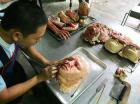
Most morgue operations place the DNA collection team near the end of the examination line after the photographs and radiographs have been taken and following the completion of the dental examination and autopsy. The chief of morgue operations or medical examiner/coroner will direct DNA samples to be taken in accordance with the scope of the investigation, as discussed earlier.
Usually the postmortem team will take a single sample from relatively intact remains to confirm or augment other identification methods.
Team members may also be required to select the best possible material from each of numerous fragmented human remains in order to provide a primary identification or the genetic basis for reassociation of body parts. Most frequently, natural disasters tend to require the former approach to sampling, whereas transportation accidents and terrorist events are more likely to have a greater need for reassociation.
The ideal DNA sample collection team includes two to three persons possessing (1) training in general morgue operations, (2) a rudimentary understanding of DNA science, and (3) thorough familiarity with human anatomy.
They must understand the criticality of the anatomical description and the unique numbering of samples, plus be able to handle a Stryker saw, tissue forceps, and scalpel with skill and safety. Odontologists and anthropologists are usually good choices for the collection team, but death investigators and emergency medical personnel are good alternatives. The trauma surrounding the event and subsequent environmental conditions will adversely affect the soft tissue first by fragmentation and later by decomposition. Although skeletal muscle is an easy sample to collect at the morgue and relatively simple to process at the laboratory, the condition of the remains may necessitate the collection of samples of bones and even teeth instead. Clumps of hair, skin flaps, and soft tissue that are predominantly composed of adipose tissue all cause additional steps in laboratory processing and should be avoided when possible.
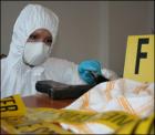
The collection team must use sterile disposable supplies when possible and wipe all surfaces and clean nondisposable gloves, instruments, and working surfaces with a 10% bleach solution between samples.
Tissue, bone, or tooth samples should be placed in a secure container without any preservative. The collection teams at the Armed Forces DNA Identification Laboratory have had greatest success using sterile 50 ml conical tubes with screw caps. Urine specimen cups may leak, glass containers could break, and small plastic bags are subject to puncture. Conical tubes with screw caps easily accommodate 5 to 25 g samples of soft tissue, bone, or tooth, do not leak, and have a smooth exterior surface for handwritten or adhesive labels. Plus, enforcing their use actually limits the amount of sample that an overly enthusiastic anthropologist, odontologist, or pathologist can submit from a single source. This reduces the long-term storage requirements of the laboratory and forces the collection team to focus on the selection of the best quality material while reducing unnecessary cutting of the remains.
The corollary to the morgue operation is the Family Assistance Center and the coordination of the collection of antemortem reference samples that come from the victim’s personal effects or family references that provide a DNA profile for comparison purposes. The family members that appear at the Family Assistance Center are not always the best genetic candidates for family references. Furthermore, most out-of-town family members may not linger long after the initial event. For these reasons, as much information regarding the victim’s genetic tree and the whereabouts of other relatives must be obtained on the first interview with the next of kin. Given the emotional displacement of family members after a disaster, predesigned forms that include family tree templates will help distraught relatives place themselves and others in the proper genetic relationship. The preferred family reference donors are listed under the section on salivary DNA.
1.4.5 Transportation and Storage
Once the collection team has verified the accuracy of the chain of custody, the samples should be transferred to the laboratory as expeditiously as possible.
Individually labeled and sealed 50 ml conical tubes are placed in heavy-duty clear zip-lock plastic bags. Depending on the size of the bags, three to five tubes can be placed in a single bag. This second layer of packaging serves two purposes. First, if leakage occurs from a tube, it limits the potentially contaminating exposure to a limited number of other samples, and it also reduces the likelihood that numerous labels will become smudged or illegible.
Second, when working with very large numbers of samples, the plastic bag simplifies the moving of evidence to and from the laboratory or in and out of storage. If precoordinated with the laboratory, collection teams can even use the bags to batch samples according to their priority. Bags of samples are best transported in a clean commercial-grade cooler. The 16-quart size or larger allows ample room for bags of ice or reusable ice packs to keep samples cold during transportation. If samples cannot be transferred to the laboratory immediately, they should be kept in a cool, dark, dry environment, preferably at -20°C. Samples may be shipped using a commercial courier, but a courier rotating directly and only between the morgue and laboratory, maintaining wireless communication with both sites, and possessing security clearances at both sites is highly desirable. This arrangement will overcome the business hour restrictions that hamper some delivery services and also will facilitate an unbroken chain of custody.
1.4.6 Data Management
The sample collection and laboratory processing portions of the DNA mass fatality response are complicated activities. But neither is more complex than the need to assemble all of the data that are generated, review them, and compare these unknown profiles with the available references, including interpreting the results and assigning a statistical weight to the conclusions. This activity becomes far more challenging depending on the number of different laboratories that are processing samples and the degree of commonality between their procedures. These issues are best addressed during the disaster planning phase. Other factors that will impact data management requirements are the typing technologies applied (autoSTR, Y-STR, mtDNA, etc.), the genealogical proximity of family references to the victims, and decisions regarding the use of partial or incomplete profiles.
Most DNA laboratories are acquiring or developing their own laboratory information management systems that aid the staff in maintaining the chain of custody and facilitate data analysis, statistical computation, and tracking both productivity and turnaround times. The variation in capability from one management system to another is quite extreme, and very few laboratories have systems that are developed specifically to handle mass fatality scenarios.
1.5 Teeth as Sources for Forensic DNA
Teeth are known to survive most postmortem events, including natural phenomena such as decomposition and autolysis, as well as environmental insults, such as water immersion, burial, and fires to as hot as 1,100°C.
Neurovascular cells of the pulp and, to a lesser extent, odontoblasts embedded in the predentin layer and trapped during mineralization in the dentinal tubules have been shown to be valuable sources of DNA evidence when other bodily tissues are degraded or missing.
Various methods are advocated to access these various regions of the tooth and effectively recover sufficient amounts of DNA from the different cell types to both minimize the potential
loss of dental structures and data and yet maximize DNA recovery.
The spectrum of various methods to access dental cells for DNA extraction are represented by the two most widely used dental DNA recovery techniques. Additionally, current research is focused on other methods to increase further the yield of DNA from degraded and compromised samples.
To conservatively access cellular material, Smith et al. advocate horizontal sectioning of the tooth near the cementoenamel junction. This retains the coronal aspect for ulterior forensic examination or historical preservation while exposing the pulp chamber and radicular pulp system, predentin, and dentin for DNA recovery.
Once the dentin has been removed from the crown, the enamel shell and any restorations are left intact. Cases that may require this approach are those that involve remains of unique cultural value or museum specimens where the destruction of the material must be minimized. Examples are investigations into the remains of Tzar Nicholas and his family, analysis of dental evidence representing members of George Washington’s extended family, and attempts to identify the putative skulls of Mozart and Fredric Schiller (unpublished data). This technique is also applied by the U.S. military in the identification of its missing in action (MIA) soldiers and in mass fatality incidents in which individual teeth are recovered and may represent the sole remains available for return to a family.
Attention is being given to other nondestructive methods, especially with respect to ancient invaluable human exhibits. Studies by Krzyżańska use a micro fluidic pump to flush cells from the tooth by rinsing the pulp system from the apical orifices through small holes in the occlusal surface.
These methods may show promise for forensic exhibits to allow DNA recovery from the pulp while preserving the tooth.
Alternatively, following the application of traditional forensic odontological identification methods, Sweet recommends that crushing or grinding the entire tooth into a fine powder can obtain the maximum quantity of DNA.
This must follow adequate documentation of the exhibit with charts, notes, photographs, and radiographs. The method allows recovery of DNA from remnants of pulpal cells as well as the embedded cells present in the hard tissues, especially in cold cases involving skeletal remains.
On a case-by-case basis, then, the laboratory must make informed decisions about the best method to employ to recover DNA. This decision should be made in concert with the forensic odontologist using his knowledge of dental histology and taking into account the presence of any identifiable morphological or restorative traits of the tooth.
1.6 Saliva and Oral Mucosa as Sources for Forensic DNA
In addit ion to being consulted on the use of teeth and the craniofacial structures as a source for human identification, the forensic odontologist should also be prepared to advise on matters concerning the biological character of oral mucosal and salivary DNA for the same reason. Salivary DNA can be recovered in a variety of scenarios and from a wide range of inanimate objects, including clothing, foods, tobacco products, oral hygiene devices, drink containers, dental prostheses, stamps, and envelopes. Laboratories will generate their own protocols for recovery of salivary DNA from personal effects, but will probably defer to the odontologist in processing a bitemark for DNA. The forensic odontologist may also be approached with questions regarding the buccal swab. As the name suggests, DNA from the oral mucosa of the inner cheek is the target of the buccal swab technique.
It is a collection method that is rapidly supplanting the older phlebotomy or finger stick techniques in which whole blood samples are collected for DNA processing.
1.6.1 Salivary DNA in Bitemark Evidence
Saliva derives from three major bilateral glands and hundreds of smaller ones scattered throughout the oropharyngeal region. Over two-thirds of the 1.0 to 1.5 L of the saliva produced daily is from the submandibular gland, whereas the parotid glands account for approximately 25% of the total volume and the sublingual glands only 5%.
The biological composition of the fluid varies between the glands, and the volume of salivary secretion at any given time of day will change according to physiological stimulation or medical issues or a pharmacological condition. But generally secretion is highest during eating.
Saliva is composed chiefly of water but also contains electrolytes, buffers, glycoproteins, antibodies, and enzymes.
The detection of one particular group of enzymes, the human α-amylases, which are responsible for initial polysaccharide digestion, is a traditional method for identifying evidentiary stains for the presence of saliva.
Although amylase is found in other bodily fluids, its very high levels in saliva are unique in that they can be as much as fifty times greater than those in other sources. Its stable biochemical
activity over time is distinctive as well.
Amylase-dependent forensic screening tests are increasingly available in the commercial sector. Some are very specific and are based on mono clonal antibody activity that focuses on human salivary α-amylase, whereas others are more general in relying on the detection of amylase activity to release a colored dye suggesting the presence of saliva.
However, all such direct-contact assays risk consumption of portions of the sample that are otherwise destined for DNA analysis. If used, consideration should be given to selecting the most informative product that requires the least volume of sample.
Visual screening for evidentiary saliva stains should be augmented by the use of alternative light sources. Although ultraviolet light is helpful in uncovering the presence of biological fluids, short-wave ultraviolet light is a potential health hazard and is also known to disrupt the DNA molecule and compromise the ability to conduct forensic DNA testing. Alternative light sources, such as lasers and high-intensity lights that can be filtered to provide a single wavelength, are probably the best for screening evidence, including skin, for the presence of saliva.
The advantage of an alternative light source in screening for salivary stains on skin is that the potential DNA sample is physically untouched and that detection can be made even in
the absence of a patterned injury.
It is the cellular component of saliva that makes it an attractive target for the DNA analyst. These cells are not secreted by the salivary glands but are incorporated into saliva as part of the shared oral environment. Specifically, oral mucosal cells are sloughed into the salivary mix through normal epithelial turnover and the activity of mastication. Additionally, white blood cells, most commonly the acute inflammatory polymorphonuclear leukocytes, arise from the crevicular fluid secondary to gingivitis. The microbial fauna that perpetually occupy the oropharyngeal spaces carry their own genomes and add to the total sum of DNA, but in the context of forensic analysis, the human-specific DNA primers do not recognize the bacterial DNA sequences.
The ability to obtain forensic DNA data from salivary evidence was well described by 1992, but it was not until Sweet et al. published the double swabbing technique in 1997 that the standard of practice for salivary DNA collection from skin was truly established.
The double swab procedure takes into consideration the need to moisten and lift the cellular component of an adherent saliva stain as well as addresses the risk that the bitemark victim’s skin will be a likely contributor to the DNA in the sample. The technique requires two sterile cotton swabs and 3 ml of sterile, distilled water.
A brief description of the procedure follows:
1. Thoroughly moisten the head of a cotton swab in sterile, distilled water.
2. Roll the head of this swab over the area of the saliva stain while using moderate pressure and a continuous circular motion.
3. Allow this first swab to air dry in a contamination-free environment for at least thirty minutes.
4. Within ten seconds of completing the first swab, roll the tip of the second, dry swab across the now moist area of the stain.
5. Use a circular motion and light pressure to absorb the moisture from the skin into the second swab.
6. Allow the second swab to air dry in a contamination-free environment for at least thirty minutes.
7. After drying, both swabs are packaged together, sealed, and marked with unique sample and case numbers.
8. The chain-of-custody document is completed and samples are submitted to the laboratory.
As an addendum to the sample collection from the victim’s skin, a DNA elimination sample should also be collected from the victim to allow the laboratory to subtract that profile from any data generated from the skin swabs.
This reference sample can be whole blood, a buccal swab (see below), or if the victim is deceased, a tissue sample taken at autopsy.
The double swab technique has a confirmed track record of success to include salivary DNA collections from a block of cheddar cheese and a body submerged in water for over five hours.
1.6.2 Oral Mucosal DNA and Buccal Swabs
A successful conclusion based on DNA analysis is dependent on the opportunity to compare a questioned profile with a known profile. An investigator has little control regarding the condition and sufficiency of questioned profiles found on the victim or at the crime scene, but a level of quality control can be exercised in obtaining the known reference profile. Whole blood samples drawn through a finger stick or veinapuncture procedure are the traditional methods in obtaining a DNA reference. Well-meaning investigators occasionally have collected head hair believing that they were procuring a simple noninvasive source of DNA. They did not realize that the laboratory processing requirements for hair samples is resource-intensive, compared to the more predictable blood sample. The taking of head hair as a DNA reference should be limited only to those cases where the questioned samples consist of shed hairs and the use of mitochondrial DNA testing is likely.
For routine casework and long-term storage, whole blood applied to a filter paper card provides the greatest amount of reference DNA by volume with the least likelihood of contamination. However, the use of a buccal swab as a rapid, noninvasive alternative to blood collection is increasingly commonplace.
Because of the anatomical specificity of the forensic odontologist’s expertise, he or she may be consulted in the collection, storage, and transportation of the buccal swab. Targets of this collection technique are the stratified squamous epithelial cells that can be rubbed from the inner cheek, although a certain amount of salivary DNA is naturally collected as well and is beneficial.
When buccal swabs are being collected to help identify a missing or unidentified family member, the laboratory will provide a list of the preferred donors according to their relationship with the victim. Exactly which family members are targeted as donors depends on the anticipated DNA typing system to be applied.
Generally, when autosomal STRs are being used, the preferred reference sources, ranked in order of desirability, include a pristine sample from the:
1. Victim (collected before the event and properly preserved)
2. Victim’s biological parents
3. Victim’s biological child and this child’s other biological parent
4. Victim’s biological child
5. Victim’s full siblings
6. Victim’s half siblings
Combinations of these references may also be extremely helpful, and the laboratory managing the case should be consulted. Recall, too, that earlier in the chapter, paternal lineage markers (Y-STRs) and maternal lineage markers (mtDNA), while not unique to the individual, may still provide inclusionary or exclusionary results. In combination with circumstantial and other forensic evidence, the results can be statistically compelling. The lineage markers also offer the potential for a reference source that spans many generations. For example, a great-grand-nephew may serve as a mtDNA reference for a victim that is missing decades earlier.
There are many commercial buccal swab collection kits on the market today.
The simplicity and painlessness of the procedure combined with the minimal training requirement for the person performing the collection has launched this method of DNA reference collection to the forefront of use.
The other advantages that buccal swabs offer over blood-based collection techniques are the ease of self-collection and greater tolerance of this procedure in children and uncooperative donors. Although the commercial market offers a series of ingenious collection devices, suitable results can be obtained with sterile cotton-tipped applicators found in most medical supply stores.
Most buccal swab collection protocols are essentially the same. Start by documenting the identity of the donor or establish a unique sample number if the identity of the donor is unknown.
Wear gloves and avoid contaminating the swabs by contact with any surface or aerosol other than that of the donor:
1. Have the donor thoroughly rinse his or her mouth with water and expectorate. Repeat.
2. Wipe one side of the buccal mucosa with 2 × 2 sterile gauze.
3. Firmly stroke the dried area of the mucosa ten times with the swab, slowly rotating the cotton tip each time. Avoid the site near the opening of Stenson’s duct if possible.
4. Repeat the process with a second swab on the contralateral cheek.
5. Allow both swabs to air dry in a contamination-free environment for at least thirty minutes.
6. Place in a prelabeled paper envelope or wrapper and seal with evidence tape.
7. Verify that the unique labeling and correct contents of the packet are documented; initial and date the seal for continuity purposes.
8. Complete the chain-of-custody form and ship to the laboratory as directed, maintaining a cool, dry, ultraviolet-light-free environment wherever possible.
Rinsing prior to DNA collection reduces the presence of food particles or debris, such as chewing tobacco, and even reduces the possibility of contamination by a nonnative DNA source.
Wiping the mucosa also eliminates adherent oral debris, such as materia alba, plaque, and microbial flora, at the site of collection. Importantly, for these same reasons, rinsing or wiping before taking an oral swab should not be done if the subject is a suspected rape victim and oral copulation may have occurred. In this particular situation, the goal is not to obtain a reference sample for a donor but rather gain biological evidence of the attacker.
The receiving laboratory will have its own protocol for processing buccal swabs. As the popularity of this collection technique grows, an increasing number of laboratories are adopting a high-throughput platform that accommodates the swab samples. In this way, hundreds or even thousands of samples containing high-quality DNA in a predictable concentration can be processed relatively quickly.
Go to Part2