Hypoglossal nerve
Hypoglossal motor nerve innervating the muscles of language, which contains sympathetic and sensory fibers. Sympathetic fibers enter the nerve to connective branches from the upper cervical node simpaticheskogo barrel. Sensory fibers coming from the lower node (gangl. infegiog).
The core of the hypoglossal nerve (nucl. n. hypoglossi) located in the medulla oblongata at the bottom of the rhomboid fossa. Lower division core stretches up to 1 11 cervical segments. Nucl. n.
hypoglossi is like a continuation of front pogov cepogo spinal processes of cell nuclei fall between the pyramids and olives of the medulla oblongata and form a common trunk.
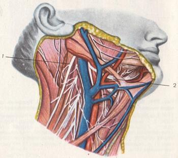
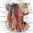
Of the cranial cavity HN couple goes through the canal hypoglossal nerve (canalis hypoglossus) of the occipital bone. Coming out of the canal, the nerve is first between the external and internal carotid arteries, and then under the rear belly of the digastric muscle descends on the lateral surface podyazychnoyazychkovoy muscles, limiting the top of the triangle
Pirogov. Then the nerve passes between chelyustnopodyazychnoy podyazychnoyazychnoy and muscle, enters into sublingual region, where the branches into terminal branches, innervating the muscles of the tongue.
The core of the hypoglossal nerve is mainly connected through korkovoyadernyh relationships with the opposite hemisphere.
Central motor neuron for the muscles of the tongue is located in the lower part of the precentral gyrus. Kortikonuklearnye fibers go through the knee internal capsule, brain stem, the bridge and the medulla oblongata. At the level of the medulla oblongata most of the fibers overlaps and fits to the nucleus n. hypoglossus opposite side.
Symptoms defeat
Any damage to the trunk or core hypoglossal nerve develops flaccid paralysis or paresis of the muscles of half of the tongue, which is accompanied by atrophy of the muscles of the tongue, fibriklyarnymi twitching. When protruding tongue deviation observed him in the direction of destruction, as healthy podborodochnoyazychnaya muscle, by subtracting the one side, and pulls the tongue forward in the opposite direction.
The defeat of the hypoglossal nerve, sometimes accompanied by a violation of speech. It is unclear, stumbling (dysarthria). When bilateral lesion occurs immobility of language and inability to articulate speech
(anarthria). Is also violated by acts of chewing and swallowing.
In addition to these symptoms in lesions of the nucleus n. hypoglossus observed dysfunction circular muscles of the mouth at the other safety features mimic muscles. This is due to the fact that the circular muscle of the mouth come hypoglossal nerve fibers in the facial nerve.
Incidences
Any damage to the internal department of the hypoglossal nerve function is disrupted only muscles of the tongue.
If the nerve is affected after the channel podyazychnogo nerve, then the process may involve fibers entering the nerve from the cervical roots, which is accompanied by dysfunction of the muscles that fix the larynx. As a result of swallowing the larynx is displaced in a healthy way.
With the defeat of the hypoglossal nerve nucleus in the medulla oblongata at the same time in the process may involve the pyramidal path, passing through the barrel. Develops alternating syndrome Jackson.
Since the core of the hypoglossal nerve is associated mainly with the opposite hemisphere, with lesions of the lower divisions precentral gyrus and korkovoyadernogo way
HN nerve paralysis occurs in the central type.
This is not observed atrophy and fibrillar tremors in the muscles of the tongue (tongue sticking out was rejected in the opposite position of the hearth).
During the processes of the internal capsule at the same time amazed kortikonuklearnye fibers and the pyramidal path, which is accompanied by the development of central paralysis and HN
VH pairs of cranial nerves and spastic hemiparesis on the opposite side. With bilateral lesions korkovoyadernyh ways, going to the IX, X and XH pair, develops so-called pseudobulbar syndrome.
TRANSLATE FROM RUSSIAN BY GOOGLE