| | |
|
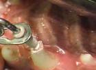
Periodontal Ligament Histology of the Teeth
06-11-2008

When embedded in the jaw in its bony crypt, the tooth is surrounded by a fbrous bag, the so-called dental follicle as has already been mentioned above, and that is not only the source of the periodontal ligament but also for the cells that form the secondary cementum and the alveolar bone.
During and after tooth eruption, the fibrous connective tissue composing the follicle transforms into the periodontal ligament that forms the connection between the tooth and the walls of the bony crypt that evolves into the alveolar socket.
At the neck of the tooth, the dent alveolar collagen us fibres of the periodontal ligament connect with those of the gingival, which are specifically known as gingival fibres. Epithelial rests of Malassez, already mentioned before, lie embedded in the collagen fibres between alveolar bone
and root surface.
The dental follicle is extremely important from a deferential diagnostic point of view. It may contain structures that are also found in a variety of odontogenic tumours and that may be mistaken as proof for presence of such a tumour by the unaware pathologist. This point will be discussed more extensively within the context of odontogenic tumours.
| |
| | |
|
|
|
|
|
| |
Comments
пїЅпїЅпїЅпїЅпїЅ:werwerкурган Гость:Hi Nick.I think you’re right; you are likely to need to do some pre-study bofere you are likely to be accepted onto a dental hygiene or other clinical course. I think the fact that you have a post graduate qualification and the fact that it is in IT is likely to work in your favour, it shows that you are intelligent, hard working, and experienced in effectively studying a particular subject. Of course having a good knowledge of computers is particularly useful in dentistry as most practices are now computerised.I would advise you to have a good look at the вЂвЂ™ and вЂвЂ™ section of my website to help you to gain a better understanding of a number of clinical roles within the dental profession, and to hopefully help you to decide on a particular career choice! Once you have decided which clinical role most interests you, you can look at the list of training institutes in the U.K. and then do some further research into preferred entry requirements, length of time it takes to complete a course etc by looking at the institutes own websites.I would also advise, once a decision has been made by yourself, that gaining some experience in a clinical environment would be a big help to you when it finally comes to applying for the course of your choice, obviously knowledge and experience are two very different things. I hope that this advice is useful and that you become successful and happy in your chosen career.Katy. Гость:X7OiXX vdqtjrhhjwob Гость:bRqucF odspgzqccyuo Гость:Shannon, Thank you for your question! We are happy to help any way we can. Unfortunately, sieiitnvsty on a tooth can be a tough thing to diagnose without taking a look. On a front tooth, it can be as simple as gum that has receded (pulled up from the tooth) that allows air to meet the root surface. The root surface is much more porous so allows anything (air, bacteria, etc) better access to the nerve of the tooth and this can, in turn, lead to sieiitnvsty. Sensitivity can also be caused by trauma to a tooth. Trauma can be anything from falling and hitting the tooth or biting into something particularly hard. Something people often don’t think about are habits. Do you have a habit of biting your nails? Holding a pen between your teeth? Any of these can cause enough trauma to the tooth that it can lead to sieiitnvsty. If it is something that continues for a week, call us (or your local dentist if you are not in our area) to have an exam and xray taken to make sure it is a simple fix! Гость:Altiazaam-informaakon found, problem solved, thanks! Гость:You write so holsteny about this. Thanks for sharing! http://fafltiu.com [url=http://pmiwdn.com]pmiwdn[/url] [link=http://grlbwolygeu.com]grlbwolygeu[/link] Гость:Can I simply say what a aid to find soemone who really knows what theyre speaking about on the internet. You positively know how to convey a difficulty to mild and make it important. More folks need to learn this and perceive this facet of the story. I cant imagine youre no more fashionable since you positively have the gift.
|
| |
Articles for theme “Histology”:
| | |
|
|
06-11-2008
The dental pulp is the soft connective tissue that forms the inner core of each tooth. It is divided in the coronal part and the radicular part depending on its position. At the root tip, the radicular part of the pulp merges with the periodontal ligament. At this site, blood vessels and nerves enter the tooth. At the interface of pulp and dentin lies a continuous row of columnar odontoblasts. They are responsible for the deposition of dentinal matrix that may be either secondary as a physiologic process continuing as long as the tooth lives, or tertiary as a reply to some insult.
|
| |
| | |
|
|
|
|
| | |
|
|
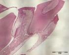
06-11-2008
Root cementum is visible as a tiny, slightly basophilic layer at the outer surface of the root part of the tooth and can be distinguished from dentin by the absence of tubuli. It obliterates the peripheral ends of the dentinal tubuli and lies adjacent to the mantle dentin. It is the product of fibroblasts from the dental follicle migrating to the root surface and differentiating into cementoblasts. Whether epithelial cells from Hertwig’s sheath contribute to initial cementum formation is controversial as has already been discussed.
|
| |
| | |
|
|
|
|
| | |
|
|
06-11-2008
Enamel content is almost 100% mineral, so it evades histologic evaluation in common paraffin sections from decalcified material. Only in immature tooth tissues, may it be seen in routinely prepared histologic slides due to a still higher proportion of organic components, where it is visible as a slightly basophilic the ready material. In this form, it may also form part of the histologic appearance of odontogenic tumours.
Examination of mature enamel requires the employment of ground sections.
|
| |
| | |
|
|
|
|
| | |
|
|
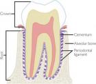
11-10-2008
Teeth consist for the major part of dentin. This material houses the dental pulp, the soft tissue core of the tooth consisting of myxoid connective tissue with blood vessels and nerves, and supports the enamel cap that covers the part of the tooth that is exposed to the oral cavity. In the root area, dentin is covered by cementum that fixes the collagenous fibres of the periodontal ligament onto the root surface. At the other side, these collagenus fibres are attached to the bone of the tooth socket and in this way, the tooth is fixed in
the jaw.
|
| |
| | |
|
|
|
|
| | |
|
|
07-06-2008
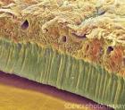
Dentin is a specialized kind of bone formed by the odontoblasts but different in the sense that it does not contain complete cells but only cellular extensions, i.e., cytoplasmic extensions from the odontoblasts. These cross the full thickness of the dentin from the odontoblastic cell body that lies at the border between dentin and dental pulp to the junction between dentin and enamel. The tiny canals that house the odontoblastic extensions are recognizable as evenly spaced tubuli. This tubular nature is the histologic hallmark for dentin, not only in teeth but also in odontogenic lesions in which the nature of each mineralized material may not be recognizable at first sight.
|
| |
| | |
|
|
|
|
|
|
|
|