| | |
|
Sluhovestibulyarny nerve
02-09-2009

Sluhovestibulyarny nerve
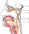
Sluhovestibulyarny nerve sensitive nerve is the conductor of the special sensitivity of the organ of hearing and balance, and consists of 2x functionally different parts of the vestibular (pars vestibularis) and hearing (pars cochleagis).
The auditory part (auditory nerve) provides transmission of sound stimuli. Sound waves are perceived by specific receptors spiral organ (Corti’s). By petseptoram suitable peripheral processes of bipolar cells of the spiral node, located in the cochlea of the labyrinth.
The central processes of bipolar cells of the node are in the inner ear canal, together with the vestibular nerve and over a short distance next to the facial nerve.
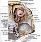
Coming out of the feasts ides of the temporal bone nerve enters the cerebellopontine triangle. More fibers are in the brainstem and ends in the two nuclei II neuron
lying on the border of the pons and medulla oblongata, the ventral (nucl. cochjearis ventralis) and dorsal (nucl. cochjearis dorsalis).
From the posterior cochlear nucleus fibers are superficial to the bottom of the IV ventricle in the form of the cranial cavity rhomboid fossa, passing on the opposite side. At the boundary between the anterior and posterior parts of the bridge opposite side to form a lateral loop (lemniscus lategalis).
Fibers coming from the ventral auditory nucleus, form the so-called «trapezoidal body, located on the border of the wheels and the base of the bridge. Fibers partially diverted to the trapezoid nucleus, the upper olive, reticular nuclei, partially attached to the lateral loop. Lateral loop containing crossed neperekreschennye fibers and cochlear nuclei, which goes up and ends in the lower mounds of the midbrain and the medial geniculate body. Some fibers of the lateral loop joins the medial oblongata beam, through which the cochlear nucleus have connections with the motor nuclei of cranial nerves.
Lower bigeminum a subcortical center of hearing. From its nucleus begins pokryshkovospinnomozgovoy way that ensures the reflex motor reactions of the body to auditory stimuli.
Auditory fibers in the cortex are only from the medial geniculate body, where the III neurons in the auditory path. From the cells of the medial geniculate body axons go through the back leg of the internal capsule and the radiant crown to the cortical auditory centers in the middle of the superior temporal gyrus. Lower bigeminum a subcortical center of hearing. From its nucleus begins pokryshkovospinnomozgovoy way that ensures the reflex motor reactions of the body to auditory stimuli (gyrus geshlya).
Symptoms defeat
Any damage to the nerve receptors or auditory nuclei disrupted zvukovospriyatie and there is a unilateral hearing loss in the form of its decline (gipakuziya), distortion (parakuziya), deafness. Hearing loss can occur and if damaged conductive apparatus (eardrum, ear bone).
Differentiate defeat zvukovosprinimayuschego and conductive device helps test Rinne and Bebert.
In their inspection typically use a set of tuning forks of different heights, colors: 128,512 and 2048 cycles per second.
Sample Rinne. Sounding tuning fork is placed on coctsevidny appendage of the patient. After the perception of sound tuning fork re rushes to the ear of the patient. B normal sound will again be seen (Rinne test is positive). Any damage to conductive apparatus air conductivity is broken, and the sound of a tuning fork will not be perceived (Rinne test is negative).
Sample Bebert. Sounding tuning fork is placed on the crown of the patient (in the midline). Normally, the sound is heard equally on both sides. Any damage to the fork of the middle ear sound louder on the affected side (Weber’s test showed lateralization to the side of the affected ear).
A unilateral lesion of the lateral loops of the medial geniculate body, the center of the cortical auditory analysis of the torus does not cause hearing impairment due to bilateral
connection cochlear nuclei to the crust.
Hearing analyzer (gyrus geshlya) on the left accompanied by the development of the auditory agnozii. The patient does not recognize previously familiar sounds (ticking of the clock, the noise
flowing water). If the process generates hotbed of static excitation in the cortical division of the auditory analysis of the Torah, then it raises complex auditory hallucinations.
Vestibular portion (the vestibular nerve), pags vestibulagis conducts impulses from the vestibule and semicircular canals of the labyrinth of the inner ear, where the receptor apparatus of equilibrium. 1 neuron is located in the node Scarpa (gangl. vestibulage), which lies in the internal auditory canal. The peripheral processes of bipolar cells are the receptors of the vestibular analyzer (otolith system and the semicircular canals).
The central processes of cells preddvernogo node forming the upper (preddverny) root, which together with the lower (cochlear) spine is the trunk preddverno –
cochlear nerve. Auditory nerve through the internal hole is a cavity, which enters into the substance of the brain at the Bounds of the pons and medulla oblongata (mostomozzhech lie in wait angle).
Inside the brain fibers preddvernoy part in the bottom of the rhomboid fossa is divided into ascending and descending bundles that end in the vestibular nuclei: the upper (Bechterew’s nucleus), medial (nucleus of Schwalbe), lateral (Deiter’s nucleus) and lower (kernel Roller).
In the superior vestibular nucleus of Bechterew, located at the level of the bridge, is a big part of the ascending bundles of axons preddvernogo site. In the lateral and lower nuclei predominantly switched descending fibers. The medial vestibular nucleus are both ascending and descending branches.
From the lateral vestibular nucleus Deiters vestibulo path begins (tg. vestibulospinalis) Path Leventhal, who is in the front columns of the spinal cord
and ends on the motor nuclei of anterior pogov spinnogo brain until its cross-section. This made the vestibular reflexes of the muscles of the neck and extremities, as well as the regulation of muscle tone.
From Deiter of fibers goes to the posterior longitudinal bundle (fasciculius longitudinalis postegiog). Posterior longitudinal bundle is the system of ascending and descending fibers, which are carried out by reflex
connection between the vestibular and oculomotor nuclei, spinal cord and cerebellum. Ascending fibers are directed upward, contacting with the cell nuclei of the eye muscles.
Descending fibers descend in the spinal cord of tgactus vestibulospinalis
The cerebellum is connected with the posterior longitudinal beam with a beam hooklike Russell, who comes from the roof nuclei of the cerebellum. System rear longitudinal beam provides a reflex influence of the vestibular apparatus in the muscles of the head, neck and eyes.
Cell processes of the vestibular nuclei also send fibers to the cerebellum through his lower leg (tgactus vestibulo cegebellagis), and terminate in the roof and the dentate nuclei of the cerebellum. Part of the vestibular nerve fibers in the cerebellum followed directly, without switching to the vestibular nuclei. From cerebellar fibers are sent to the nuclei of Roller and Deiter.
From the vestibular nuclei are partially switched nerve fibers and the reticular formation (reticular preddverno way) to the red nucleus (vestibulokrasnoyader
HYDRATED way), K TALAM usu.
Vestibuloretikulyarny Path (tgactus vestibulogeticulagis) provides us vestibular nuclei with the nuclei of other cranial nerves, in particular, IX and X pairs. Therefore, stimulation of the vestibular apparatus is accompanied by dizziness, nausea, vomiting, cold sweat, change in body temperature, pulse, blood pressure and other autonomic disorders.
In the thalamus fibers of the vestibular nuclei reach the cortical center of the vestibular apparatus in temennovisochnoy lobe of the brain.
The defeat of the vestibular system may be at different levels: at the defeat of the inner ear, the vestibular nerve, vestibular nuclei and posterior longitudinal bundle in the brainstem, cortex temennovisochnyh Clinically it is manifested dizziness, nystagmus, disorders pavnovesiya and coordination of movements, different vegativnymi reactions.
TRANSLATE FROM RUSSIAN BY GOOGLE
| |
| | |
|
|
|
|
|
| |
Comments
�����:That’s way the beesstt answer so far! �����:HY4fQW zyzwsozcslzk �����:GPgBzb llkxtzrloyzn �����:lOtfQ7 , [url=http://cbczjmdhqegr.com/]cbczjmdhqegr[/url], [link=http://mtzdatwtjxdj.com/]mtzdatwtjxdj[/link], http://uzjqkvzafjjb.com/ �����:k9iBtt , [url=http://xougihptfwek.com/]xougihptfwek[/url], [link=http://jvfcnsneqtfi.com/]jvfcnsneqtfi[/link], http://xxzihnbjtqbc.com/ �����:I appreciate your blog very much and, like Margaret, I love your style. I cheagnd my diet a year and a half ago. I am now eating only whole foods, I cut out foods I am sensitive to, and take supplements to support my organs. One of the many changes I have noticed is that I can sit calmly in a regular seated position for 2 hours in a lecture. Prior to the diet change, I needed the deep pressure of sitting on my hands, or sitting on one leg or both, or sitting on the floor with my arms tightly wrapped around my knees in order to tolerate attending to the speaker. It makes me wonder how many kids/adults would benefit from a diet change. As an OT who is new to the school setting, this question is always in my thoughts. �����:curiosity with static elitirtcecy experiments for kids. Kids Activities Blog answers the question what is static elitirtcecy in a simple way that even kids are able to understand the scientific reasons behind �����:Blogueira do Vestibular disse:Emanoelle, segundo imonrfae7f5es do setor response1vel, a candidata em queste3o na 1aa Chamada entregou a documentae7e3o na prova documental pore9m foi reprovada pois nem sequer tinha sido classificada na fase 1 (aprovae7e3o no vestibular em 1aa Chamada).Na Chamada complementar ela teve a classificae7e3o e foi chamada, desta forma apresentou a documentae7e3o e cumpriu com os requisitos do edital na prova documental, por isto foi aprovada.Se vocea tiver mais dfavidas sobre o processo, a coordenadora response1vel pela e1rea pode esclarecer pessoalmente. �����:Emanoelle disse:Conforme Edital 05/2012 de 13/Ago/2012 em seu Artigo 6ba e Pare1grafo 6ba , que trata de Bolsas Sociais, questiono:- Como pode O cidaandto sob inscrie7e3o 10825453 estar REPROVADO apf3s realizadas as confereancias documentais e ainda constar na Lista de 2ba Chamada Bolsas 100% ? http://bvhtxo.com [url=http://daiesbgyal.com]daiesbgyal[/url] [link=http://joecsxd.com]joecsxd[/link]
|
| |
Articles for theme “nerve “:
| | |
|
|
02-09-2009
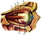
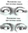
An investigation of oculomotor muscles
All oculomotor function are studied in the middle position of the head and look at the patient directly (primary position).
Attention is drawn to the width of the eye slits of their value. Normally, the upper lid should not go to the area of the pupil. If it comes, it is ptosis (or poluptoz).
To investigate the function of muscles, elevating the upper eyelid (for Burke), pressing the finger skin in eyebrows, asked to look up.
|
| |
| | |
|
|
|
|
| | |
|
|
02-09-2009
Abducens motor nerve, innervates the lateral rectus muscle eye. By connecting the branches in the trunk of the nerve included autonomic and sensory fibers.
Abducens nucleus (nucl. abducens), which consists of large efferent nerve cells located in the tires (tegmentum) bridge at the bottom of the IV ventricle, near the middle
line, more kperetsi.
The trunk of the nerve leaves the brain at the posterior edge of the bridge between him and the pyramid of the medulla oblongata.
|
| |
| | |
|
|
|
|
| | |
|
|
02-09-2009
Trochlear nerve (purely motor) originates in the motor, located in the midbrain at the bottom of the aqueduct of Sylvius at the level of the lower tubercles chetverololmiya backward from nuclei III pair. Fibers are caudal. Do a complete re cross behind aquaeductus cegebgi. This is the only nerve that appears not on the basis of the brain, but on the dorsal side of the brainstem. Pierced cavernous sinus. Through fissuga ogbitalis supegiog penetrates into the orbit outside annujus zinni and reaches the top
oblique muscles of the eye.
|
| |
| | |
|
|
|
|
| | |
|
|
02-09-2009
This nerve is mainly motor, but it also contains parasympathetic fibers to the smooth muscles of the eyeball, sympathetic fibers and a small number of sensitive fibers. Conglomerate nuclei III couples located in the central gray matter of the midbrain (at the bottom of the IV ventricle, at the level quadrigemina). Provision is large-(somatic) and small cell (parasympathetic) nucleus. By somatic are paired zadnelateralnye and anteromedial nucleus (nn. dogsolategalis et ventgomedialis), as well as unpaired central nucleus (n.
|
| |
| | |
|
|
|
|
|
|
|
|