|
|
|
|
The clinical picture of fractures of the upper jaw
03-09-2009
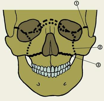 Land increased strength of the upper jaw depending on the structure of bone tissue related to its formation in phylogenesis. Strong space correspond to points of ossification, the weak – the intermediate lines. Plots of reduced strength are placed along the joints connecting it with the other bones of the facial skeleton and the bones forming the base of the skull. These sites often pass through neognestrelnogo fracture of the upper jaw. This may explain the fact (along with other factors – venue traumatic force, its direction relative to the buttresses), which is most often a fracture line is not strictly within the anatomical boundaries of the upper jaw, but extends to the neighbor, the associated bone. Therefore, the clinic faced not only with a broken most of the upper jaw, as with the «breaking» it with lots of other bones of the face and skull base. This is what determines the diversity of clinical manifestations, severity of injury and various outcomes of the upper jaw.
Le Fort (1901) experimentally identified and described the different types of fractures of the upper jaw. Currently, more likely to use his proposed classification. In accordance with the order of their descriptions identified by the upper (Le RDF I), middle (Le Fort II) and lower (Le Fort III) types of fractures. In the literature, they often do not lead to the author's version. For example, Schroeder (1916) Type II described as type I, type I – as a type II; Vasmund (1927) – type III as type I, II – as III, a I – as type II. We are in presenting the material adhere to the classification of the author, according to which the upper jaw fractures are bilateral, and the lines they are symmetrical. However, clinical experience and literature data indicate that this is not always the case.
Fractures of the upper jaw up from 2 to 5% of fractures faces. Their reason – heavy mechanical injury: motor vehicle accident, fall victim to the height or drop heavy objects on the face, kicked in the face, etc. These fractures may be accompanied by a brain injury. A broken upper jaw can be shifted backward in the direction of the applied force; down – because of its own weight fragments and a medial pterygium traction and proper chewing muscles (at the turn of type I and II). The rear of the maxilla displaced mostly down, due to the thrust of the medial pterygium muscle (see the function of muscles of the back). In the case of a traumatic force direction in oblique projection otlomok might shift further in the opposite direction of the applied force.
|
|
|
|
|
|
|
|
|
|
| |
Articles for theme “upper jaw”:
|
|
|
|
|
03-09-2009
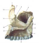
The upper jaw bone of steam, is connected with the zygomatic, frontal, nasal, Lattice, sphenoid, lacrimal bone. In her distinguished body and four sprouts: frontal, alveolar, palatal and zygomatic. In the body of the upper jaw is aeriferous maxillary sinus, the walls of which are represented by thin lamellae of the compact substance. There are four body surface of the upper jaw: the front, infratemporal, orbital, nasal. The front surface, fades anterior, limited infraorbital edge (top), skuloalveolyarnym crest and the zygomatic process (laterally), alveolar bone (below), the nasal notch (medial).
|
|
|
|
|
|
|
|
|
|
|
|
|
|