|
The upper jaw
03-09-2009
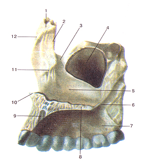
The upper jaw bone of steam, is connected with the zygomatic, frontal, nasal, Lattice, sphenoid, lacrimal bone. In her distinguished body and four sprouts: frontal, alveolar, palatal and zygomatic. In the body of the upper jaw is aeriferous maxillary sinus, the walls of which are represented by thin lamellae of the compact substance. There are four body surface of the upper jaw: the front, infratemporal, orbital, nasal.
The front surface, fades anterior, limited infraorbital edge (top), skuloalveolyarnym crest and the zygomatic process (laterally), alveolar bone (below), the nasal notch (medial). Below infraorbital region located infraorbital foramen, for infraorbitale, which goes through the terminal branches of the same name of the nerve and blood vessels. Infratemporal surface, fades infratemporalis, forms the border infratemporal and pterygopalatine pits and brought to bump the upper jaw. It is attached to the oblique head of the lateral muscles pterygium. Hillock upper jaw has a 3.4 aperture, through which the thickness of the bone tissue includes rear upper alveolar branches involved in the formation of posterior upper dental plexus.
The orbital surface, facies orbitalis, participates in the formation of the lower wall of the orbit and forms the infraorbital region. In the posterior part of it, together with the orbital edge of large sphenoid wing creates a lower orbital fissure, fissura orbitalis inferior. Through her into the orbit enters infraorbital nerve, n. infraorbitalis, – a branch of the maxillary nerve. The latter is located in the infraorbital groove and infraorbital canal. These anatomical formation located on the orbital surface of the body of the upper jaw. At the bottom wall of the channel are small front and medium-sized upper alveolar openings, – foramina alveolaria superiora anteriora et media. They are a small bone tubules, extending to the roots of incisors, canines and small molars. They are vessels and nerves to these teeth. The medial edge of the orbital surface is connected with the lacrimal bone, orbital plate of ethmoid bone and the orbital process of palatine bone. Sometimes it produces cells that are directly adjacent to the cells of the labyrinth ethmoid.
Nasal surface, facies nasalis, is connected with the perpendicular plate of palatine bone, the lower turbinate and ethmoid HOOK offshoot. At the surface between the lower and middle shells is opening the maxillary sinus – maxillary cleft, hiatus maxillaris. Anterior to the cleft is nasolacrimal duct, opening into the nasal cavity. In the formation of its participating lacrimal bone and lacrimal outgrowth of the lower turbinate. Posterior to the maxillary cleft palatine canal is a large, educated palatine bone and the wing-offshoot sphenoid bone.
Frontal processus, processus frontalis, the inner edge is connected to the nasal bone, the top – with the bow of the frontal bone, posterior – with lacrimal bone. It consists mainly of compact substance. He is able to withstand the compressive load from the bottom up to 470-500 kg, which is much more power pressure developed chewing muscles.
Zygomatic process, processus zygomaticus, connects rough surface with the zygomatic bone. Lower down from it toward the hole of the first molar is skuloalveolyarny crest. Zygomatic process and consists mainly of compact substance.
Palatine processus, processus palatinus, is a horizontal plate of bone. Forward and outward, it goes into alveolar bone, the inner surface is connected with the palatine offshoot of the opposite side, rear – with a horizontal plate of palatine bone. The inner edge of the appendix is located nasal crest, crista nasalis, which connects to the cartilaginous part of the nasal septum. The medial edge of the appendix from the palatal surface thickened. On the upper surface of the palatal sprouts on the side of the nasal ridge located incisal opening, which leads to the incisive canal, canalis incisivus. In the front 2 / 3 appendage consists of compact and spongy substance. In the posterior third of the sponge is missing, and in this department he is much thinner than the front. Palatine processus noted increased durability.
Alveolar bone, processus alveolaris, is a continuation of the body of the upper jaw downwards and consists of outer and inner plates of compact substance. Between them there are sponge. The outer plate is thinner than the inner, at the level of premolars – thicker than the front of teeth. Behind the third molar of a large outer and inner plates converge, forming alveolar hill, tuber alveolaris. Edge sprouts, limbus alveolaris, has 8 tooth holes (the alveoli) for the roots of teeth. The latter are separated by bone mezhalveolyarnymi partitions. The shape and size lunochek conform to the shape and size of the roots of teeth.
Maxillary sinus – the largest of the paranasal sinuses. It can spread in the alveolar, the zygomatic, frontal and palatine processes. In sinus distinguish upper, lower, medial, anterolateral, zadnelateralnuyu wall, covered with mucous membrane. The upper wall of the maxillary sinus is separated from the orbit. At a great distance, she presented a compact substance, its thickness from 0,7 to 1,2 mm. It thickens from infraorbital region and zygomatic process. The bottom wall of the infraorbital canal and the furrows of the same name, passing here, is very thin.
The bottom wall of the sinus – the bottom – has a form of troughs, where it joins the medial, anterolateral and zadnelateralnaya wall. The bottom of the trough or flat, or represented papulose protrusion above the roots of teeth. The thickness of the compact disc that separates the bottom of the maxillary sinus from the second hole of a large molar tooth, may not exceed 0,3 mm.
The medial wall consists entirely of compact substance and borders the nasal cavity. Thick (about 3 mm) is in the anteroinferior corner of the smallest (1,7-2,2 mm) – in the middle of its lower edge. Behind becomes zadnelateralnuyu wall. In place of this transition, it is very thin. The front of the medial wall passes into the anterolateral where thickens. In verhnezadnem division wall is a hole – maxillary cleft (hiatus maxillaris), connecting the bosom of middle nasal passage.
Anterolateral wall of the sinus in the canine fossa consists entirely of compact substance and in this place is very thin (0,2-0,25 mm). It thickens with distance from the fovea, reaching a greater thickness (up to 6.4 mm) at the infraorbital edge of orbit. We alveolar, zygomatic, frontal appendages inferolateral edge of the orbit is spongy substance. In the anterolateral wall are several alveolar ducts, where the nerve Stalks and blood vessels to the front teeth and premolars.
Zadnelateralnaya wall provided compact disc, which forks in place of transition in the zygomatic and alveolar bone. There is a spongy substance. In the upper section is thinner than that near the alveolar bone. In the thicker walls are rear alveolar ducts, which are nerve Stalks are going to the big molars. Features of the structure of the upper jaw determine the place of least resistance, the impact force, which determines the nature of the fracture. Therefore, we must again emphasize that the upper jaw is involved in the formation of the orbit, nose and mouth and is connected with the zygomatic, palatine, frontal, nasal, lacrimal, Lattice, sphenoid bone. The frontal, ethmoid and sphenoid bones, together with the temporal form the anterior and middle cranial fossa.
The walls of the maxillary sinus are thin bony plates. Nevertheless, the upper jaw is able to withstand significant mechanical loads. This is due to the fact that the trabeculae of the spongy substance are predominantly vertical type structure, and the compact substance – the thickening in certain areas, or buttresses. Their four.
▲ frontonasal buttress meets the front group of teeth. Rests on several thickened wall alveoli of the canines, Located along the edge of the frontal nasal openings and the appendix of the upper jaw to the fore the appendix of the frontal bone.
▲ Skuloalveolyarny – starts from the second premolar, first and second molars. Continues skuloalveolyarnomu ridge toward the body of the zygomatic bone and zygomatic process of frontal bone. By the zygomatic arch pressure is transmitted to the temporal bone. Is the most powerful buttresses, perceiving pressure occurring in the above-mentioned teeth.
▲ Meckel – begins at the posterior alveolar process and corresponds to the rising ground of the upper jaw and wing-appendage sphenoid bone. It also involved the formation of pyramidal appendage palatine bone, which fills the clipping pterygium pterygoid spike.
▲ palatine palatine buttress formed offshoot of the upper jaw and is represented by two longitudinal troughs, reaching the bottom of the nose. In the area of the nasal notch is connected to the fronto-nasal buttresses, which in turn is connected with skuloalveolyarnym in the upper and lower edges of the orbit. Alveolar bone unites skuloalveolyarny, pterygopalatine and palatine buttresses.
The above anatomical features determine the stability of the upper jaw to chewing pressure and its ability to withstand significant mechanical stress.
| |
