| | |
|
|
08-09-2009
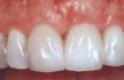
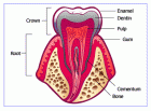
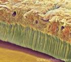
The enamel of the tooth are the hardest tissue in the body, due to its high content of inorganic substances (95%). In addition, water is present in the enamel, which is located in the free and bound state. Organic matter is in the form of lamellae. enamel tufts and belief gene. Matrix of enamel is a macromolecular Comp Lex formed fibrillar proteins and Ca. The mineral part of enamel prisms are brought united mi in bundles, which run from the enamel-dentinal connections to the surface of the tooth.
|
|
|
| |
|
|
|
|
|
|
| |
|
|
| |
|
06-11-2008
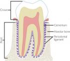
When embedded in the jaw in its bony crypt, the tooth is surrounded by a fbrous bag, the so-called dental follicle as has already been mentioned above, and that is not only the source of the periodontal ligament but also for the cells that form the secondary cementum and the alveolar bone. During and after tooth eruption, the fibrous connective tissue composing the follicle transforms into the periodontal ligament that forms the connection between the tooth and the walls of the bony crypt that evolves into the alveolar socket.
|
|
|
| |
|
|
|
|
|
|
| |
|
|
| |
|
06-11-2008
The dental pulp is the soft connective tissue that forms the inner core of each tooth. It is divided in the coronal part and the radicular part depending on its position. At the root tip, the radicular part of the pulp merges with the periodontal ligament. At this site, blood vessels and nerves enter the tooth. At the interface of pulp and dentin lies a continuous row of columnar odontoblasts. They are responsible for the deposition of dentinal matrix that may be either secondary as a physiologic process continuing as long as the tooth lives, or tertiary as a reply to some insult.
|
|
|
| |
|
|
|
|
|
|
| |
|
|
| |
|
06-11-2008
Root cementum is visible as a tiny, slightly basophilic layer at the outer surface of the root part of the tooth and can be distinguished from dentin by the absence of tubuli. It obliterates the peripheral ends of the dentinal tubuli and lies adjacent to the mantle dentin. It is the product of fibroblasts from the dental follicle migrating to the root surface and differentiating into cementoblasts. Whether epithelial cells from Hertwig’s sheath contribute to initial cementum formation is controversial as has already been discussed.
|
|
|
| |
|
|
|
|
|
|
| |
|
|
| |
|
06-11-2008
Enamel content is almost 100% mineral, so it evades histologic evaluation in common paraffin sections from decalcified material. Only in immature tooth tissues, may it be seen in routinely prepared histologic slides due to a still higher proportion of organic components, where it is visible as a slightly basophilic the ready material. In this form, it may also form part of the histologic appearance of odontogenic tumours. Examination of mature enamel requires the employment of ground sections.
|
|
|
| |
|
|
|
|
|
|
| |
|
|
| |
|
11-10-2008
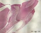
Teeth consist for the major part of dentin. This material houses the dental pulp, the soft tissue core of the tooth consisting of myxoid connective tissue with blood vessels and nerves, and supports the enamel cap that covers the part of the tooth that is exposed to the oral cavity. In the root area, dentin is covered by cementum that fixes the collagenous fibres of the periodontal ligament onto the root surface. At the other side, these collagenus fibres are attached to the bone of the tooth socket and in this way, the tooth is fixed in the jaw.
|
|
|
| |
|
|
|
|
|
|
| |
|
|
| |
|
07-06-2008
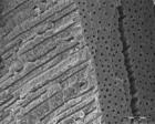
Dentin is a specialized kind of bone formed by the odontoblasts but different in the sense that it does not contain complete cells but only cellular extensions, i.e., cytoplasmic extensions from the odontoblasts. These cross the full thickness of the dentin from the odontoblastic cell body that lies at the border between dentin and dental pulp to the junction between dentin and enamel. The tiny canals that house the odontoblastic extensions are recognizable as evenly spaced tubuli. This tubular nature is the histologic hallmark for dentin, not only in teeth but also in odontogenic lesions in which the nature of each mineralized material may not be recognizable at first sight.
|
|
|
| |
|
|
|
|
|
|
|
|
|