|
Articles for theme “Nerve”:
| |
|
|
| |
|
02-09-2009
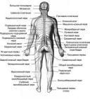
Anatomical features The autonomic nervous system Vegetative (autonomous) nervous system of the nervous system that regulates the body of the internal organs, glands of internal and external secretion, blood and lymph vessels, trophism tissue and homeostasis. Functioning on a subconscious level, she quickly and continuously respond to disturbances that threaten the permanence of the internal environment. For me nowadays, the term autonomic nervous system originates from the work of Bichat (Bishat, 1800).
|
|
|
| |
|
|
|
|
|
|
| |
|
|
| |
|
02-09-2009
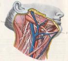
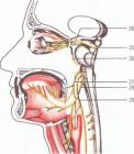
Hypoglossal nerve Hypoglossal motor nerve innervating the muscles of language, which contains sympathetic and sensory fibers. Sympathetic fibers enter the nerve to connective branches from the upper cervical node simpaticheskogo barrel. Sensory fibers coming from the lower node (gangl. infegiog). The core of the hypoglossal nerve (nucl. n. hypoglossi) located in the medulla oblongata at the bottom of the rhomboid fossa. Lower division core stretches up to 1 11 cervical segments.
|
|
|
| |
|
|
|
|
|
|
| |
|
|
| |
|
02-09-2009
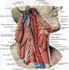
Accessory nerve Accessory nerve motor. Spinal nucleus dobavochnogo nerve occurs in the lower section of the medulla oblongata, and gray matter of the spinal cord at the level of C1C5. Aksony these cells form a 6 – 7 rootlets, which extend to the lateral surface of the spinal cord and merge into a common trunk. Barrel accessory nerve enters the cavity Th Turnip through the foramen magnum. Spinal part of accessory nerve also contains efferent fibers from the cervical segments of the brain and spinal afferent fibers nodes received through with front and rear mozgovymi spinal roots.
|
|
|
| |
|
|
|
|
|
|
| |
|
|
| |
|
02-09-2009
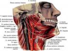
Vagus nerve The vagus nerve is a mixed nerve. Innervates the organs of the neck, chest cavity, digestive tract, organs retroperitoneal space. Has the following core: three common core with nerve IX (nucl. alae cinegea, nucl. Ambiguus, nucl. Tgactus solitagius) and own parasympathetic nucleus nucl. dogsalis n. vagi. The vagus nerve contains motor, sensory and autonomic (parasympathetic) fibers. Motor fibers coming from the motor neurons of the dual-core to the striated muscle of the soft palate, pharynx, larynx and upper esophagus.
|
|
|
| |
|
|
|
|
|
|
| |
|
|
| |
|
02-09-2009
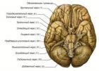
Glossopharyngeal nerve Pair of cranial nerves IX (p. glossophagyngeus) mixed nerve contains motor, sensory and parasympathetic (secretory) fiber, has 4 cores, which are located in the posterior part of the medulla oblongata. The double nucleus, nucl. ambiguus (in common with a pair of X), located in the middle part of the medulla oblongata, in front and lateral nucleus hypoglossal nerve. Axons of cells form the core motor branch of the glossopharyngeal nerve, which innervates the muscle only shiloglotochnuyu (ie stylopharingeus).
|
|
|
| |
|
|
|
|
|
|
| |
|
|
| |
|
02-09-2009
Sluhovestibulyarny nerve Sluhovestibulyarny nerve sensitive nerve is the conductor of the special sensitivity of the organ of hearing and balance, and consists of 2x functionally different parts of the vestibular (pars vestibularis) and hearing (pars cochleagis). The auditory part (auditory nerve) provides transmission of sound stimuli. Sound waves are perceived by specific receptors spiral organ (Corti’s). By petseptoram suitable peripheral processes of bipolar cells of the spiral node, located in the cochlea of the labyrinth.
|
|
|
| |
|
|
|
|
|
|
| |
|
|
| |
|
02-09-2009
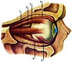
An investigation of oculomotor muscles All oculomotor function are studied in the middle position of the head and look at the patient directly (primary position). Attention is drawn to the width of the eye slits of their value. Normally, the upper lid should not go to the area of the pupil. If it comes, it is ptosis (or poluptoz). To investigate the function of muscles, elevating the upper eyelid (for Burke), pressing the finger skin in eyebrows, asked to look up.
|
|
|
| |
|
|
|
|
|
|
| |
|
|
| |
|
02-09-2009
Abducens motor nerve, innervates the lateral rectus muscle eye. By connecting the branches in the trunk of the nerve included autonomic and sensory fibers. Abducens nucleus (nucl. abducens), which consists of large efferent nerve cells located in the tires (tegmentum) bridge at the bottom of the IV ventricle, near the middle line, more kperetsi. The trunk of the nerve leaves the brain at the posterior edge of the bridge between him and the pyramid of the medulla oblongata.
|
|
|
| |
|
|
|
|
|
|
| |
|
|
| |
|
02-09-2009
Trochlear nerve (purely motor) originates in the motor, located in the midbrain at the bottom of the aqueduct of Sylvius at the level of the lower tubercles chetverololmiya backward from nuclei III pair. Fibers are caudal. Do a complete re cross behind aquaeductus cegebgi. This is the only nerve that appears not on the basis of the brain, but on the dorsal side of the brainstem. Pierced cavernous sinus. Through fissuga ogbitalis supegiog penetrates into the orbit outside annujus zinni and reaches the top oblique muscles of the eye.
|
|
|
| |
|
|
|
|
|
|
| |
|
|
| |
|
02-09-2009
This nerve is mainly motor, but it also contains parasympathetic fibers to the smooth muscles of the eyeball, sympathetic fibers and a small number of sensitive fibers. Conglomerate nuclei III couples located in the central gray matter of the midbrain (at the bottom of the IV ventricle, at the level quadrigemina). Provision is large-(somatic) and small cell (parasympathetic) nucleus. By somatic are paired zadnelateralnye and anteromedial nucleus (nn. dogsolategalis et ventgomedialis), as well as unpaired central nucleus (n.
|
|
|
| |
|
|
|
|
|
|
|